Growth Factor Binding Capacity
10 likes | 123 Views
This study examines the incorporation of heparin-modified gelatin microparticles (MPs) into embryoid bodies (EBs) to enhance differentiation and growth factor retention of embryonic stem cells (ESCs). Through controlled aggregation, these microparticles are designed to sequester morphogens released during differentiation, thereby engineering a favorable microenvironment. Results indicate that the presence of heparin-modified MPs significantly affects gene expression and growth factor levels in EBs, highlighting the potential of this approach for directed differentiation of ESCs and improved understanding of embryonic development.

Growth Factor Binding Capacity
E N D
Presentation Transcript
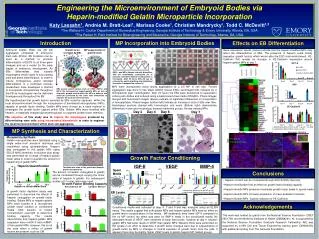
Introduction Effects on EB Differentiation Conclusions Acknowledgements Engineering the Microenvironment of Embryoid Bodies via Heparin-modified Gelatin Microparticle Incorporation Katy Lassahn1, Andrés M. Bratt-Leal1, Marissa Cooke1, Christian Mandrycky1, Todd C. McDevitt1,2 1The Wallace H. Coulter Department of Biomedical Engineering, Georgia Institute of Technology & Emory University, Atlanta, GA, USA 2The Parker H. Petit Institute for Bioengineering and Bioscience, Georgia Institute of Technology, Atlanta, GA, USA MP Incorporation into Embryoid Bodies Embryoid bodies (EBs) are 3D cell aggregates comprised of embryonic stem cells (ESCs). EB formation can be used as a method to promote differentiation of ESCS to all three germ lineages and as a model for the early stages of embryonic development. As EBs differentiatethey secrete morphogenswhich signal to surrounding cells and affect differentiation. In order to induce homogenous and/or directed differentiation in a controlled manner, researchers have developed a method to incorporate microparticles throughout the EB so morphogens can be delivered homogenously throughout the EB. Gene expression results (below) indicate that the heparin-modified MPs may affect the differentiation of EBs. The presence of heparin could locally sequester growth factors which would alter the ESC microenvironment. VE-Cadherin IHC reveals an increase in VE-Cadherin expression around heparin-gelatin MPs at day 7. Growth factor secretion by EBs MP sequestration of growth factor A B Time Differentiation C ESCs and MPs (labeled red with AlexaFluor 594) are sequentially centrifuged (A) to promote controlled material incorporation in microwells. After 24 hours EBs (B) can be removed by gentle pipetting and maintained thereafter in suspension culture(C). MPs were incorporated using forced aggregation at a 2:5 MP to cell ratio. Forced aggregation was done in two steps withD3 mouse ESCs centrifuged first, followed by a microparticle layer centrifugation. After 24 hours the EBs were transferred to suspension culture petri dishes and cultured using a basal serum-free media (ESGRO). Phase images merged with fluorescent images (above) indicate no significant differences in incorporation of microparticles. Phase images (bottom left) indicate an increase in size of EBs over time. Histological sections stained with hemotoxylin and eosin (Bottom right) demonstrate differences in size and morphology between treatment groups. Arrows indicate MPs. EBs secrete growth factors such as VEGF, BMP-4, and IGF-II. By incorporating heparin-gelatin MPs we aim to sequester growth factors within the MPsin order to use molecules produced by the stem cells themselves to engineer the local microenvironment. We hypothesized that growth factors secreted by EBs could be captured within the local microenvironment through the incorporation of biomaterial microparticles (MPs), capable of growth factor binding. Gelatin MPs were chosen as a base material to investigate the growth factor capture within EBs. Gelatin MPs were modified with heparin, a negatively charged glycosominoglycan, to augment growth factor retention. ** The objective of this study was to capture the morphogensproduced by differentiating stem cells using incorporated biomaterials in order to engineer the local microenvironment within stem cell aggregates. ** ** Day 7 *=Significantly different than No MP (*=p<.05, **p<.005) †=significantly different than Gelatin (p<.05) MP Synthesis and Characterization Microparticle Synthesis Gelatin microparticles were fabricated using a single water-in-oil emulsion technique and crosslinkedusing glutaraldehyde. Heparin was conjugated to the gelatin MPs using EDC/S-NHS chemistry. A toluidine blue assay was performed for a range of heparin:gelatin molar ratios in order to quantify the amount of heparin/mg of gelatin MPs. ** ** Day 2 Day 4 Day 8 EBs were cultured in serum-free media. No MP Heparin-Gelatin Gelatin VE-Cadherin Hoechst Microparticles Growth Factor Conditioning No MP IGF-II VEGF BMP-4 Spent Media The amount of heparin conjugated to gelatin can be modulated through varying the molar ratios of heparin to gelatin. For subsequent studies a 5:1 molar ration was used. Gelatin • Heparin content can be modulated through EDC/S-NHS chemistry • Heparin modification has an effect on growth factor binding capacity • Heparin-Gelatin MPs presence modulates growth factor levels in spend media • Heparin-Gelatin MPs increase expression of meso-endoderm markers • Heparin-Gelatin MPs localize induction of VE-Cadherin Growth Factor Binding Capacity Gelatin Heparin-Gelatin Heparin-Gelatin *=Significantly different than No MP (p<.05) †=significantly different than Gelatin (p<.05) A growth factor depletion assay was performed to determine the effects of heparin conjugation on growth factor binding. Gelatin MPs or heparin-gelatin MPs were soaked in a recombinant growth factor solution or conditioned media (CM) solution of known concentration overnight to determine binding capacity. The results demonstrate that heparin-gelatin MPs sequester more rmIGF-II and rmBMP-4 than gelatin MPs, however, this is not the case when a milieu of growth factors are present such as CM. EB Lysate Conditioned media was collected at days 5, 7 and 9 and was analyzed using an ELISA assay. The results suggest that both gelatin MPs and heparin-gelatin MPs have an effect on growth factor concentrations in the media. MP treatments have lower IGF-II compared to the no MP control. No effect was seen for BMP-4 levels in the conditioned media. No detectable levels of VEGF were observed at early time points, however at day 9, heparin-gelatin MPs had lower levels of VEGF than gelatin MPs. Western Blots were performed on EB Lysate in an effort to determine if growth factor changes were due to sequestration of growth factor by MPs or changes in normal secretion of growth factor from the cells. It appears there may be slightly higher VEGF levels in gelatin-heparin MP treated groups. Toluidine blue stain of the gelatin and heparin-gelatin microparticles. This work was funded by grants from the National Science Foundation (CBET 0651739) and the National Institutes of Health (GM088291). KL is supported by the National Science Foundation Graduate Research Fellowship. ABL was supported by a NIH Cell and Tissue Engineering training grant (GM008433) with additional funding from The Goizueta Foundation.