Download
1 / 58
600 likes | 1.07k Views
Learn about upper and lower airway obstructions in children, including signs, symptoms, and etiologies. Understand the importance of prompt recognition and management to prevent respiratory distress. Explore common congenital and acquired causes, such as infections and traumatic etiologies, with an emphasis on differentiating inspiratory and expiratory obstructions.

E N D
AIRWAY OBSTRUCTION IN CHILDREN PEDIATRIC RESPIROLOGY CHILD HEALTH DEPT. GADJAH MADA UNIVERSITY
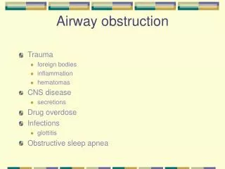
INTRODUCTION • AIRWAY OBSTRUCTION : • * Upper Airway obstruction • * Lower Airway obstruction
Stridor • Stridor, which is derived from Greek, meaning “creaking,” is caused by rapid turbulent flow through a narrowed airway. • The sound generated depends on the degree of constriction and the localization of the obstruction. • Observation of the child often offers the best clue to localization, before a cold stethoscope ever touches the chest.
Lokasi lesi stridor • Supraglottic lesions, such as epiglottitis, present with inspiratory stridor, a prolonged inspiratory phase • Glottic lesions also lead to high-pitched inspiratory stridor and a weak or hoarse voice. • Subglottic lesions cause expiratory stridor with a normal voice and a brassy cough
Signs and symptoms of respiratory distress • Tachycardia • Tachypnea 0-2 months RR P 60x/min 2-12 months RR P 50x/min 1-5 years RR P 40x/min • Suprasternal retractions indicate more severe obstruction than intercostal and subcostal retractions.
impending respiratory failure include: - marked retractions, - decr. or absent breath sounds, - increasing tachycardia, - decreasing respiratory effort or rate, - decreasing stridor, - a worried or unsettled appearance. - Ominous signs are decr. Level of conc., extreme pallor, head- bobbing with each breath, and decr. Heart rate. Cyanosis is an extremely late sign in upper airway obstruction
ETIOLOGY • Airway obstruction may be congenital or • acquired • Congenital : • The result of anomalies of development • that impact upon airway function • Congenital etiologies must be considered • in young infants, and less common • causes are tumors, and edema secondary • to severe allergic reaction.
Acquired : • The consequences of some process - infection, - trauma, - neoplasia, - inhalation of foreign body
Traumatic etiologies must also be considered, including foreign bodies, • external trauma to the neck, burns, and • iatrogenia (ex. Postintubation) • Upper airway obstruction is a common • cause of pediatric emergency department • visits, accounting for approximately 15% • of all critically ill patients.
• Infectious etiologies account for 90% of these, • with viral croup accounting for 80%. • Epiglottis accounts for 5% of severe • cases. • • Other significant causes include other • infections etiologies • (bacterial tracheitis, tonsillar pathology, • mononucleosis, and diphtheria)
Inspiratory obstruction = extrathorade • The vibratory sound produced by inspiratory obstruction is heard during inspiration, is usually monophonic, • high pitched as in croup or • low to medium pitched as in snoring resulting from adenotonsillar hypertrophy.
Congenital malformations • Nasal, nasopharyngeal, and oropharyngeal malformations • - Retrognathia (Pierre Robin syndrome) • - Nasal, choanal, or nasopharyngeal • stenosis; tumor, mass Craniopharyngioma. • Anterior encephalocele • Teratoma • - Adenotonsillar hypertrophy • - Obesity or redundant pharyngeal tissue • - Hypotonia (e.g., Down syndrome) • - Oral cavity or pharyngeal tumor • - Lingual tumor : Lingual thyroid tumor, Hemangioma • - Neck masses : bronchial cleft cyst, Cystic hygroma
Laryngeal or subglottic airway malformations • - Laryngomalacia • - Paralyzed vocal cords • - Laryngeal or arytenoid cysts • - Laryngocele • - Subglottic stenosis • - Subglottic hemangioma • Infection • - Nasal, nasopharyngeal, and oropharyngeal infection. • Tonsillitis and peritonsillar abscess • Sublingual abscess (Ludwig’s angina) • Retropharyngeal abscess
Allergy and asthma • - Anaphylactoid reaction to food or inhalant • - Vocal cord dysfunction • Metabolic problem • - Hypocalcemia or hypomagnesemia • Acquired tumor (rare)
Expiratory obstruction = intrathoracic The vibratory sound produced by this obstruction is best heard on expiration and may be focal or monophonic and of low to medium pitch or may be diffuse or polyphonic and of medium to high pitch. Congenital malformations Tracheobronchial tree malformations Tracheobronchomalacia Primary (focal or diffuse) tracheobronchomalacia Tracheobronchomalacia secondary to compression by tumor (focal).
Expiratory obstruction = intrathoracic • Congenital malformations– • Tracheostenosis • VATER (vertebral defects, imperforate anus, tracheo esophangeal fistula, radial and renal dysplasia), association • Complete tracheal rings • Vascular compression (ring or sling) • - Aberrant subclavian vein • - Pulmonary artery sling ( aberrant left pulmonary artery) • - Right-sided thoracic aorta with left ductus arteriosus • - Left-sided thoracic aorta with right ductus arteriosus • - Double aortic arch. • Dilated cardiac chamber or dilated pulmonary artery with compression
Infection - Intrinsic airway narrowing Bronchitis Bronchiolitis Laryngotracheobronchitis Bacterial tracheitis Bronchiectasis Cystic fibrosis Juvenile respiratory papillomatosis (late) - Extrinsic airway compression Mycobacterial or fungal infection with lymph node enlargement. Infection or congenital foregut malformations, cysts Lung abscess.
Foreign body or aspiration • - Gastroesophageal reflux with bronchitis • - Foreign body in airway • - Foreign body in esophagus • Trauma • - Tracheobronchial burns or scalds • - Tracheobronchial injury (blunt or penetrating) • Allergy and asthma • - Anaphylactoid reaction to food or inhalant • - Asthma with inflammation or bronchospasm • Autoimmune disease • - Bronchiolitis obliterans after lung or bone • marrow transplant • - Idiopathic bronchiolitis obliterans
Tumor - Primary airway narrowing > Hamartoma > Benign tumors (e.g., lipoma, chondroma, myoblastoma) > Malignant tumor Bronchial adenoma Bronchogenic carcinoma Sarcoma - Extrinsic airway compression > Hodgkin’s lymphoma > T cell lymphoproliferative disease with mediastinal mass > Sarcoma Pulmonary edema
Croup • • Laryngotracheobronchitis • • Most common cause of infectious • acute upper airway obstruction. • • 10% of children with croup require admission, • 1-5% require intubation,
Pathophysiology • Transmitted via the respiratory route. • Initial port of entry is the nose and nasopharynx • Begins with a prodrome of a few days of mild URI with nasal congestion, sore throat and cough. • As the infection spreads distally, so does the edema. • A hoarse voice and harsh, brassy, bark like cough (barking like a seal) • Stridor usually develops at night
• Viral etiologies include parainfluenza virus type 1, influenza, respiratory syncytial virus (RSV), rhinoviruses and measles. • Mean age of affected patients is 18 months, with a slight male predominance, and there is a seasonal increase in cases in autumn and early winter.
Croup • May have elevated temperature. • Drooling is uncommon. • May have mild expiratory wheezing • Inspiratory stridor at rest with nasal flaring, suprasternal and intercostal retractions. • Poor air entry • Lethargy + agitation = HYPOXIA • Dehydration
Treatment of Croup • Humidified air or oxygen Provides H2O and penetrates the area of inflammation and add moisture to the mucosa. The viscosity of the secretions in the trachea which facilitates clearance of the airways. • Steroids are controversial • Albuterol treatment 2.5mg in 3ml NS. • Racemic epinephrine., is indicated for children with stridor rest or marked increase in work of breathing. It has been shown to decrease airway obstruction. Max. effect is seen in 30 min. with rebound in 2 hours.
Epiglottitis • Also known as supraglottitis • • First described in 1878, was • thought to be disease of • adults. “angina epiglottidea • anterior” • • 60% male dominance
Epiglottitis • Occurs in children from 3-7 yrs in age with only 4% under the age of 1. • Hemophilus influenzae (bacterial infection) is the most common etiology. (some viruses, allergic reactions and physical and thermal injuries can play a part also) • 1985 – vaccine….but things mutate.
Pathophysiology • Local invasion of the epiglottis occurs followed by bacteremia. • The epiglottis, aryepiglottic folds, false vocal cords, and supraglottic structures become inflamed and edematous, leading to narrowed airway and respiratory compromise. • Inspiratory airway occlusion often occurs prior to total occlusion from supraglottic edema.
Pathophysiology con’t…. • Ingesting hot liquids may develop of epiglottitis. • Scald burns to the face. • Caustic ingestions • Foreign bodies • Inhalation injuries • Sidestream exposure to crack cocaine • Burns from a crack cocaine pipe screen filter.
The evidence….. • Swollen epiglottis (the thumb sign) • Thickened aryepiglottic folds • Obliteration of the vallecula
Signs and symptoms • Very sudden onset and progresses rapidly • Muffled voice or cry (in croup it is more hoarse) • Minimal cough • Sore throat, fever, hoarseness • Drooling caused by difficulty swallowing saliva • Intercostal muscle retractions • Noisy, high-pitched, squeaky inhalations • Purple skin and nails • Odd head posture. (sniffing position), tripod position
Why do children with epiglottitis have airway obstruction? • Fatigue • Laryngospasm • Progressive swelling of the supraglottic structures • Pooled secretions
Treatment of epiglottitis • DO NOT AGITATE THE CHILD IN ANY WAY • Airway mgmt. • Administer high flow humidified oxygen in order to obtain maximal alveoli oxygen saturation. • If there is an obstruction – ventilation. • Position of comfort • Run like hell………………
Croup • Voice – hoarse • Cough – barking • Fever – yes • Saliva – minimal • Neck swelling – little • Begins – slowly • Season – autumn • Time – evening/night Epiglottitis • Voice – muffled • Cough – usually none • Fever – yes • Saliva – lots • Neck swelling – lots • Begins – suddenly • Season – all year • Time – all day
Remember…………… • Failure to manage the airway is the leading cause of preventable deaths in children.
General Treatment • Airway, airway, airway • Supplemental oxygen • Position of comfort • REMEMBER – almost all children with an U.A.O. can be bag-valve-mask ventilated, and this should always be tried first in a respiratory failure situation. • Advanced airway management, IV, cardiac monitor, Pulse ox, etc.
General Treatment con’t….. • Always have a smaller ET tube readily available because of the possibility of significant airway edema. • Watch for aspiration
DEFINITION • Chronic inflammatory disease of the air way • associated with - airway hyperresponsiveness • - air flow limitation • - respiratory symptoms • Air way inflammation • * Acute bronchoconstriction • * Swelling of the air way • * Chronic mucous plug formation • * Air way remodelling
ASTHMA Allergen, viruses, Chemical sensitiser, emotions, weather /seasonal changes, pollution? Inflammation eosinophilic bronchitis Triggers Symptoms cough, wheeze,dispneu, chest tightness Airway hyper responsiveness
ASTHMA MANAGEMENT • Key components: assesment • management • Assesment of asthma severity • - Severity of asthma attack • - The episode of asthma (Classification of Asthma Severity )