Download

1 / 32
320 likes | 576 Views
Membrane-induced bundling of actin filaments. Nature Physics 4 , 789-793 (2008). Actin protrusions emerge from dendritic actin networkss. GUVs (prepared in sucrose solution) ~15um G-actin/ Arp2/3/ N-WASP/ GTP- γ S Actin filament protrusions formed within first few minutes.

E N D
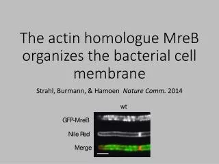
Membrane-induced bundling of actin filaments Nature Physics 4, 789-793 (2008)
Actin protrusions emerge from dendritic actin networkss GUVs (prepared in sucrose solution) ~15um G-actin/ Arp2/3/ N-WASP/ GTP- γ S Actin filament protrusions formed within first few minutes Membrane protrusions are supported by actin filaments
AF488 GUV ~ 40um Arp2/3 uniformly distributed w/o visible side chains Filament barbed ends are concentrated at the tip of protrusions (CP) Monomer addition occurred primarily at its tip AF555 10um
Elongation of a thin protrusion (phase-contrast) Length of protrusion: 1-25um (n>1000) Filament remain straight w/o lateral fluctuations Initial rate: 1um min-1 Protrusion growth initially quickly but slows down over time.
Photobleaching on actin along the filament protrusion Monomer addition does not occur along the thin actin filament protrusions
Too simple to be true? • RESEMBLANCE to the cellular filopodia • Elongation occurs at the tip of the protrusion • Protrusions lack the dendritic architecture • But the filament protrusion formation don’t require bundling or tip-complex proteins • The reasons against the in vitro filament protrusions experiment. • dendritic actin network geometry and resistance of the membrane to deformation • Interplay between actin network and membrane mechanics.
Estimation on the membrane-mediated forces between protruding filaments Energy of membrane σ, surface tension; κ, bending rigidity; H, curvature Actin filaments s, arc length along the filament; L, fixed contour length of the filament; kBT, unit of thermal energy Lp, rigidity of the polymer Geometric constraints Stable state of two merged filaments at d<10nm d=D-2x Lprot =L- L0 -y(x, L)
Membrane deformation and filament bending L-L0, protrusion length LFprot, force limited length LBprot, Branch-limited maximum protrusion length Membrane elasticity is sufficient to bring together the tips of two nearby nascent protrusions.
Balance of polymerization force and membrane rigidity Kinetic limit of filament length due to branching leads to a decrease in the range of attraction for low membrane tension
Membrane tension and filament bundling Bundling of filaments is largely tension-independent at high membrane tension, but becomes less effective at low membrane tension
Stability of thin actin filament protrusions against Euler buckling Ebundle, bundling energy of the filaments Emem, membrane energy Lmem, length of the membrane tube fmem, membrane resistance force Total energy of the deformation is lower for a straight protrusion than for a buckled protrusion
A balance between polymerization force and membrane resistance 1) Local deformation merge of deformation Bundling enough filaments to overcome the membrane resistance to tube formation, proto-filament elongate without further physical constraint ==> Membrane induced alignment of actin filaments could facilitate formation of filopodia in cells.
Mutant in the N-terminus of ParA1scoe • Double labeling (YFP-ParA and CyPet-ParB) • Co-localization of the ParA foci with ParB • Photobleach experiment • ParA1 dynamic movement in/on nucleoid • Bacteria two Hybrid • Role of ParA1-NTD in protein-protein interaction
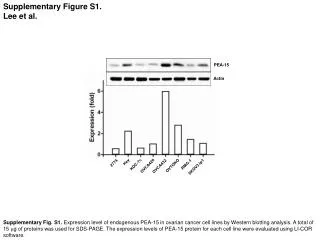
Mutants in the N-terminal domain YFP DAPI merge DIC R8E R19E R26E R19E+R26E Q29E V14G R31E
Localization in the presence of ParB Plac-yfp::parA1-parB_silent mutation on parS merge YFP DAPI DIC merge Plac-yfp::parA1-B YFP DAPI DIC The bright spots could be the nucleation of ParA1 on the parS-ParB complex
FRAP, data processing Raw data ROI1 ROI Tot BG Tot ROI3 ROI2 ROI1 BG Correction X normalization factor [ROI(t)-BG(t)]/[Tot(t)-BG(t)] X [Tot(t0)-BG(t0)]/[ROI(t0)-BG(t0)]
FRAP, kinetic plot and fitting AIM, fitting Molecular dynamics, I=I0 - I1x e -t/τ IΔ Fitting I1 I0 t1/2 I1 IΔ IΔ IΔ not not I1 + IΔ I0 I1 + IΔ I0
Difference between the growth stages Bleach regions (ROI selection) Inter or intra nucleoid movement Different chemical interactions between cytoplasm and nucleoid localization Number of molecules (is this meaningful in an inducible system?)
considerations Thin cellular extensions 63X 1.4NA oil immersion objective Maximize the difference between bleaching and imaging mode 100-fold higher Short wavelengths, high-intensity illuminations antioxidants Bleach on the optical axis Pinhole/ low NA objective Lower zooms to monitor neighboring control cells Good spatial and temporal resolution are achieved at high zooms Timing bleaching time < 1/10 T1/2 ; data collection >10-50X T1/2 Offset slightly greater than zero Gain few or no pixels are saturated Increase the effective dynamic range of measurements 12-bit images (4096 gray values)
Results Intranucleoid exchange of ParA1scoe is much faster than inter-nucleoid exchange The immobile fraction in the not separated nucleoid is comparatively larger than those in separated nucleodi. Two phases of ParA1 movement
Inter-nucleoid movement Intra-nucleoid movement
Interaction of ParA1scoe-NTD with ParA1scoe, ParA1scoeNC, and ParA1scoe-NTD (assayed with B2H) 1) There is no ParA1scoe-NTD interaction with other parts of ParA1scoe detectable in B2H experiment. 2) Intermolecular association between ParA1scoe is very strong.
NEXT PROJECT • parSA1B on miniF-sopABC--functional assay, test on plasmid stability • B2H experiments --learn the interplay between ParA, ParANC, and ParB --assess on the effect of mutants on ParA1 association • In vitro experiments --learn the interplay between parS-ParA-ParB • Gel shift/ microMacs/ Biacore