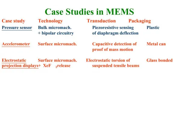
Case Studies in Infrahyoid Neck
810 likes | 1.2k Views
Case Studies in Infrahyoid Neck. Nicholas A. Koontz, M.D. Neuroradiology Fellow, University of Utah. Financial Disclosures. But first…. Please direct your smart phone, tablet, or laptop’s browser to:. Objectives. Review Infrahyoid Neck Anatomy Deep Spaces Nodal Stations

Case Studies in Infrahyoid Neck
E N D
Presentation Transcript
Case Studies in Infrahyoid Neck Nicholas A. Koontz, M.D. Neuroradiology Fellow, University of Utah
But first… • Please direct your smart phone, tablet, or laptop’s browser to:
Objectives • Review Infrahyoid Neck Anatomy • Deep Spaces • Nodal Stations • Cases, Cases, Cases • Tackle challenging cases • Develop an appropriate differential diagnosis • Identify useful discriminators • Multiple choice questions
Anatomic Spaces of Infrahyoid Neck Visceral Space Carotid Space Retropharyngeal Space Perivertebral Space Posterior Cervical Space
Case 1 • 65 year-old woman with neck pain, palpable lump
Differential Diagnosis Differentiated Thyroid Ca Medullary Thyroid Ca Anaplastic Thyroid Ca Thyroid NHL Multinodular Goiter
Most Likely Diagnosis Differentiated Thyroid Ca (DTCa) Age & Sex Ill-defined Infiltrating, invasive Mixed solid/cystic Intra-thyroidal Calcs Intra-thyroidal Intra-nodal Adenopathy Some solid Some cystic Punctate calcs
Question 1 • Which of the following is a TRUE statement? • A. Follicular is the most common subtype of DTCa • B. Hematogenous spread is more commonly associated with Papillary carcinoma • C. The peak incidence of DTCa is seen in women in the third or fourth decade • D. Rising free T4 is a clinical marker for disease recurrence
Question 1 • Which of the following is a TRUE statement? • A. Follicular is the most common subtype of DTCa • B. Hematogenous spread is more commonly associated with Papillary carcinoma • C. The peak incidence of DTCa is seen in women in the third or fourth decade • D. Rising free T4 is a clinical marker for disease recurrence
4) 30 year-old-woman, adenoma Magnified Cor CECT of LN
Case 2 • 55 year-old-woman with right neck mass, cough
Differential Diagnosis H&N SCCa Metastatic Nodes Systemic Nodal Metastases Thyroid Ca Metastatic Nodes HL or NHL Nodes Granulomatous Lymph Nodes Reactive Adenopathy
Most LikelyDiagnosis Systemic Nodal Mets Infrahyoid (level IV) location H&N primary SCCa more commonly levels II & III Non-calcified Sarcoid, DTCa often Ca++ Central low-density/necrosis HL, NHL, & reactive nodes usually solid, but can be low-density
Use Everything at Your Disposal “I’ll tell you right now – that ain’t normal.” -- Rick Wiggins
Question 2 • Which of the following is MOST suggestive of systemic nodal metastases in the neck? • A. Enlarged suprahyoid (level I or II) node • B. Enlarged left supraclavicular lymph node • C. Centrally necrotic lymph node • D. Calcification within an enlarged cervical node
Question 2 • Which of the following is MOST suggestive of systemic nodal metastases in the neck? • A. Enlarged suprahyoid (level I or II) node • B. Enlarged left supraclavicular lymph node • C. Centrally necrotic lymph node • D. Calcification within an enlarged cervical node
HL with“Signal” Node AKA Virchow node Isolated left supraclavicular adenopathy look to the chest & abdomen for primary Most HL patients present with neck nodes Concurrent mediastinal nodes common Rarely extranodal H&N disease M > F Peak incidence in mid-20s
Question 3 • Which of the following is a TRUE statement? • A. HL is more common than NHL • B. Extranodal disease favors HL over NHL • C. Imaging can reliably differentiate NHL from HL • D. HL has an earlier peak incidence than NHL
Question 3 • Which of the following is a TRUE statement? • A. HL is more common than NHL • B. Extranodal disease favors HL over NHL • C. Imaging can reliably differentiate NHL from HL • D. HL has an earlier peak incidence than NHL
Case 4 • 55-year-old woman with known thyroid nodules, reportedly benign – surveillance US
Longitudinal Transverse Power Doppler
Differential Diagnosis Congenital lesion Lymphatic malformation Venolymphatic malformation Venous malformation 3rdBranchial cleft cyst Neurofibroma Schwannoma Malignant Lymph node Carotid artery Pseudoaneurysm
Most LikelyDiagnosis Congenital lesion Lymphatic malformation Benign, circumscribed No flow on US Demonstrably separate from IJV and CCA Venolymphatic malformation Possible, but would have essentially no venous component Why not a NST?
Carotid Space Nerve Sheath Tumor Pros Cons Echogenicity Lack of vascularity • Location • Size • Morphology • Low Density Image c/o Lauren Ladd, M.D.
Question 4 • Which of the following is a FALSE statement? • A. Most lymphatic malformations are diagnosed before age 2 • B. Lymphatic malformations can be acquired • C. Lymphatic malformations have no malignant potential • D. Microcystic lymphatic malformations are less likely to recur than macrocysticmalformations
Question 4 • Which of the following is a FALSE statement? • A. Most lymphatic malformations are diagnosed before age 2 • B. Lymphatic malformations can be acquired • C. Lymphatic malformations have no malignant potential • D. Microcystic lymphatic malformations are less likely to recur than macrocysticmalformations
Case 5 • 25-year-old man with enlarging neck mass, recent URI