Micro-hematocrit
130 likes | 653 Views
Learn about microhematocrit testing in medical technology, including the principle, specimen requirements, procedure, reference ranges, sources of error, and the Rule of Three for quick visual checks. Ensure accurate results with proper techniques.

Micro-hematocrit
E N D
Presentation Transcript
Micro-hematocrit Introduction To Medical Technology - Lab 13 -
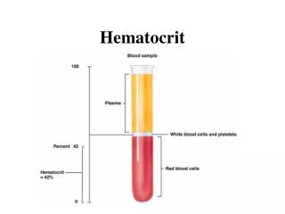
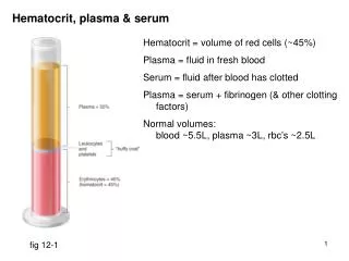
Micro-hematocrit (Packed Cell Volume Of Whole Blood) • Hematocrit is defined as the volume occupied by erythrocytes in a given volume of blood and is usually expressed as a percentage of the volume of the whole blood sample. The hematocrit may also be referred to as Packed Cell Volume (PCV).
Principle: The hematocrit is usually determined by spinning a blood-filled capillary tube in a centrifuge. Specimen: Venous blood anticoagulated with EDTA or capillary blood collected directly into heparinized capillary tubes can be used. Specimens should be centrifuged within 6 hours of collection. Hemolyzed samples cannot be used for testing.
Reagents, supplies, and equipment: Capillary tubes, heparinized for finger sticks (red tip) or plain for anticoagulated blood (blue tip) -75 mm long Clay-type tube sealant Microhematocrit centrifuge Microhematocrit reader Kimwipes or gauze
Fill two capillary tubes approximately three quarters full with blood anti-coagulated with EDTA or heparin. Alternatively, blood for heparinized capillary tubes may be collected by capillary puncture. Wipe any excess blood from the outside of the tube. Seal the end of the tube with the non colored ring with nonabsorbent clay. Balance the tubes in the centrifuge with the clay ends facing the outside away from the center, touching the rubber gasket. Procedure:
Tighten the head cover on the centrifuge and close the top. Activate the centrifuge for 5 minutes between 10,000 and 15,000 rpm (see comments). Do not use the brake to stop the centrifuge. Determine the HCT by using a micro-hematocrit reading device. Read the level of RBC packing. Do not include the buffy coat (leukocytes and platelets) when reading. The values of the two Hct’s should agree within 2% (0.02).
Hematocrite Reader Reference Ranges: • Newborn 53-65% • Infant/child 30-43% • Adult male 42-52% • Adult female 37-47%
Sources Of Error And Comments • Improper sealing of the capillary tube causes a decreased Hct reading as a result of loss of blood during centrifugation. a higher number of erythrocytes are lost in relation to the plasma. • An increased amount of anti-coagulant decreases the Hct reading as a result of erythrocyte shrinking. • A decreased or increased result may occur if the specimen was not properly mixed.
The time and speed of the centrifugation and the time when the results are read are very important. Insufficient centrifugation. Time for complete packing should be determined for each centrifuge and rechecked at regular intervals. • The micro-hematocrit centrifuge should never be forced to stop by applying pressure to the metal cover plate. This will cause the RBC layer to “sling” forward and results in a falsely elevated value. • If too much time elapses between when the centrifuge stops and the capillary tube is removed, the red cells can begin to settle out and cause a false reading of the hematocrit.
The buffy coat of the specimen should not be included in the Hct reading, because its inclusion falsely elevates the result. • A decrease or increase in the readings may be seen if the micro-hematocrit reader is not used properly. • A number of disorders such as: • Sickle cell anemia • Macrocytic anemia's • Hypochromic anemia's • Spherocytosis • Thalassemia may cause plasma to be trapped in the erythrocytes even if the procedure was performed properly.
The trapping of the plasma causes the microhematocrit to be 1-3% (0.01-0.03 L/L) higher than that obtained on automated instruments, which calculate the Hct and are unaffected the trapped plasma. • A temporarily low Hct reading may result immediately after a blood loss, because plasma is replaced faster than erythrocytes. • Proper specimen collection is an important consideration. The introduction if interstitial fluid from a skin puncture causes decreased Hct readings.
The Rule Of Three • When specimens are analyzed, by either automated or manual methods, a quick visual check of the results of Hb and Hct can be done by applying the “rule of three.” This rule applies only to specimens that have normal erythrocytes. • The value of the Hct should be three times the value of the Hb ±3. • Example: The following results are obtained from a patient: • Hb = 12.0g/dL Hb (12)x3=36; Hct=0.36L/L • Hct = 0.36L/L • Acceptable range for the Hct would be 0.33-0.39L/L