Download
1 / 74
740 likes | 759 Views
Learn about different types of CNS trauma, including focal and diffuse injuries, primary and secondary trauma, and closed versus open trauma. Explore definitions, epidemiology, craniocerebral injuries, and more.

E N D
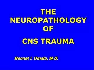
THE NEUROPATHOLOGY OF CNS TRAUMA Bennet I. Omalu, M.D.
OUTLINE: section 5 1. Definitions 2. Epidemiology 3. Craniocerebral injuries: Scalp, Skull, Intracranial cavity, Brain, Spinal cord, diffuse axonal injury 4.Sequelae of trauma 5. Non-accidental trauma in childhood
DEFINITIONS • CNS trauma : Injury or damage to living CNS tissue caused by an extrinsic agent or force by either direct or indirect mechanisms comprising: a. direct blunt force trauma b. direct penetrating force trauma c. indirect translational force trauma d. indirect asphyxiational trauma Synonyms: Traumatic brain injury, Craniocerebral injury, Head trauma
DEFINITIONS CNS trauma can be subdivided into: • Focal trauma: focal distribution of trauma • Diffuse trauma: diffuse distribution of trauma • Primary trauma: immediate and direct CNS response to trauma • Secondary trauma: delayed physiologic CNS response to trauma • Closed CNS trauma : dura mater is not disrupted by trauma • Open CNS trauma : dura mater is disrupted by trauma
DEFINITIONS • Direct blunt force trauma • CNS injuries that are due to the impact of a blunt object/ surface on the head/ body or vice versa • e.g. : blow on the head with a baseball bat fall from a tenth floor balcony
DEFINITIONS Direct penetrating force trauma • CNS injuries that are due to the impact of a sharp object on the head/ body resulting in penetration/ perforation of CNS tissue e.g.: gunshot wound of the head stab wound of the orbital cavity
DEFINITIONS Indirect translational force trauma • CNS injuries that are due to accelerating, decelerating and rotational kinetic energy, which are translated into shearing forces that disrupt CNS tissue and vessels. • e.g.: ‘whiplash’/ shaken baby syndrome
DEFINITIONS Indirect asphyxiational trauma • CNS injuries that are due to agents or mechanisms of trauma that will sufficiently reduce blood and/ or oxygen supply to the brain to result in reversible or irreversible neuronal metabolic deficits • e.g.: suicidal hanging smoke inhalation/ carbon monoxide intoxication
EPIDEMIOLOGY • 500, 000 - 750, 000 cases of CNS trauma per year • 10% are fatal • 30 - 50% are moderate/ severe • 5 - 10% result in residual deficits • 150/ 100, 000 population exhibit sequelae of CNS trauma
EPIDEMIOLOGY • Leading cause of death in people under 45 years of age • Accounts for 1% of all deaths • Accounts for 30% of deaths from trauma • Accounts for 50% of deaths due to road traffic accidents
CRANIOCEREBRAL INJURIES: SCALP • Abrasions of the scalp • Contusions of the scalp • Scalp hemorrhages, subcutaneous • Scalp hemorrhages, subgaleal • Lacerations of the scalp • Incised wounds of the scalp • Gunshot wounds of entrance and exit
Abrasions of the scalp • Scraping and removal of the superficial layers of the skin (epidermis and/ or upper dermis) • Commonly a product of blunt force impacts • Eccentric and marginal tags of epidermis on an abrasion indicate direction of impact • Patterned abrasions: imprints of the surface of impacting object on the skin
Contusions of the scalp • Hemorrhage into the skin or underlying soft tissue without breaching the skin which can manifest as: 1. Subcutaneous or intra-galeal hemorrhage: Hemorrhage into the fibro-adipose tissue of the scalp 2. Subgaleal hemorrhage: Hemorrhage below the epicranial aponeurosis (galea aponeurotica)
Subgaleal hemorrhage Subcutaneous scalp hemorrhage
Contusions of the scalp • Commonly a product of crushing impacts that rupture bloodvessels • Rate of degeneration of extravasated red blood cells and heme can be used to date scalp contusions
Laceration of the scalp • A tear of the fibroadipose and aponeurotic scalp due to perpendicular or glancing blunt force impact • Edges are usually undermined and accompanied by marginal abrasions • Tissue bridges consisting of nerves, connective tissue and blood vessels connect the margins of lacerations
Incised/ stab wounds of the scalp • An incised wound is a cut that is longer than it is deep • Produced by a sharp-edged object drawn over the scalp • Wound edges are straight without marginal abrasions or tissue bridges • A stab wound is deeper than its length on the skin • Produced by penetration of a pointed object into the depth of the scalp/ head
Gunshot wounds of entrance • Typically a circular perforating defect with loss of tissue • +/- rim of marginal abrasions, +/-radiating marginal lacerations • Contact wounds: +/- muzzle imprint, +/- soot deposits • (range: < 15cm) • Close range wounds: punctate abrasions (powder stippling/ tattooing) around the wound due to particles of propellant (range: 30 - 45 cm) • Underlying perforating defect in skull shows inward bevelling of margins
Gunshot wounds of exit • Typically an ellipsoid or stellate perforating defect without loss of tissue • Marginal abrasions are usually absent • +/- radiating marginal lacerations • Soot deposits and powder stippling are absent • Underlying perforating defect in skull shows outward bevelling of margins
Gunshot wounds of entrance Gunshot wound of exit
CRANIOCEREBRAL INJURIES: SKULL • Fractures of the cranium • Non-depressed linear fractures • Depressed Circular and curvilinear fractures • Comminuted displaced fractures • Sutural diastasis • Fractures of base of skull • Longitudinal/ axial fractures • Transverse fractures • Ring fractures • Blow out fractures
Non-depressed linear fractures of skull • Typically produced by blunt force impacts • Multiple fracture lines radiate from epicenter of point of impact • Fracture lines are oriented in direction of impacting force • Typically occurs when a mobile head impacts a stationary flat surface • Puppe’s rule for sequencing of injuries: the course of a linear fracture will be interrupted by an antecedent fracture line
Depressed circular and curvilinear fractures • Typically produced by focal blunt force impacts of a mobile object on stationary head • Inward displacement of outer and inner bone plate fragments • May exhibit pattern of concentric ripples of fracture line
Comminuted displaced fractures • Multiple fracture lines and fragmentation of bone typically produced by severe blunt force impacts and shot gun wounds of the head • mosaic/ spider’s web fracture pattern or • Pond fracture pattern
Sutural diastasis • Separation of the cranial sutures most commonly the sagittal suture • Typical due to severe blunt force impacts • Occurs more commonly in children • A marker of non-accidental mechanisms of trauma
Base of skull: Longitudinal fractures • Front to back linear fractures • Can divide entire skull base into two halves, right and left • Produced by severe blunt impacts on the face, forehead or occiput
Base of skull: Transverse fractures • Side to side linear fractures • Can divide entire skull base into two halves, front and back • Hinge fracture: complete transverse fracture in middle cranial fossa • Produced by severe blunt impacts on either side of the head or the chin
Base of skull: Ring fractures • Circumferential fracture around foramen magnum • Separates rim of foramen magnum from remainder of skull base • Produced in a fall from significant height • Severe blunt impacts on the feet or buttocks on landing • Vertebral column is driven into the skull
Base of skull: ‘Blow-out’ fractures • Comminuted fractures of the orbital plates of frontal bone • Mechanism not well established • May involve contre-coup impacts of frontal lobes on orbital plates • May involve violent increases in intracranial pressure as seen in shotgun wounds i.e. ‘blow-out’
CRANIOCEREBRAL INJURIES: TRAUMATIC INTRACRANIAL HEMORRHAGES • Epidural (extra-dural, subperiosteal) hemorrhage • Subdural hemorrhage • Subarachnoid hemorrhage • Intraventricular hemorrhage
Epidural hemorrhage • Occurs in 10 - 15% of severe CNS trauma • Usually occurs in the presence of skull fracture accompanied by dural separation and tearing of dural vessels • Rare in the elderly because of markedly adherent dura to cranium • Commonly occurs in children without skull fracture
Epidural hemorrhage • Most common scenario: lateral hemispheric location fracture of squamous temporal bone Laceration of middle meningeal artery • Associated with a lucid interval due delayed onset of bleeding caused by spasm of lacerated artery
Subdural hemorrhage (SDH) • Usually occurs without a skull fracture • Commonly occurs as a result of translational shearing forces on the bridging subdural veins • May occur without significant blunt force impact • proclivity in the elderly due to cerebral atrophy and accentuated subdural space
Subdural hemorrhage (SDH) • Acute SDH: symptom onset < 24 hrs • Subacute SDH: symptom onset 24 hrs - 7 days • Chronic SDH: symptom onset > 7 days • SDH become organized with time • Age of SDH can be estimated with sequence of histologic changes
Histologic dating of Subdural hemorrhage Time Dural surface (outer membrane) Arachnoid surface (Inner membrane) 24 hrs Thin fibrin layer Thin fibrin layer 2-3 days Sparse mononuclear cells in fibrin Rare mononuclear cells in fibrin Rare fibroblast 4-5 days Sparse fibroblasts Rare fibroblasts Rare hemosiderophage 5-10 days 3 - 5 fibroblast layers Rare hemosiderophages neovascularization: capillaries Sparse fibroblasts Sparse hemosiderophages 10-20 days 10 - 20 fibroblast layers 14-28 days: 2-4 fibroblast layers Prominent capillaries No capillaries Some hemosiderophages 21-28 days Collagenization and fibrous membrane formation mths. - yrs: fibrous membrane: inner is less than half thickness of outer membrane
Subarachnoid hemorrhage (SAH) • Traumatic SAH commonly occurs around the cerebral fissures and basal cisterns of the brain • May accompany cerebral contusions • Acute ethanol intoxication and heavy use of alcohol carry an increased risk of SAH following trivial blunt impact
Subarachnoid hemorrhage (SAH) • Fatal basal SAH can follow severe blunt impacts on the face and forehead; and severe hyperextension of the head and neck • The basilar and/ or vertebral arteries are lacerated in such a scenario • Remote SAH is associated with xanthochromia (hemosiderin deposits) of the leptomeninges
Intraventricular hemorrhage (IVH) • Traumatic IVH as a sole finding is due to blunt impacts of the head on a hard surface during a fall • Usually arterial in origin • Usually accompanies SAH, extensive contusions of the brain and penetrating injuries of the brain • Traumatic porencephaly: extensive contusions and lacerations of the cerebrum leading to a free communication between lateral ventricle and subarachnoid space
CRANIOCEREBRAL INJURIES: BRAIN • Contusions • Lacerations • Transections • Pulpefaction • Diffuse axonal injury • Diffuse vascular injury
Contusions of brain Causes • Blunt impacts of the brain on the inner skull plate due to unidirectional inertia of the brain to violent motion of the skull • Tissue shearing forces at the moment of severe blunt impacts • Intra-cranial expansile cavitation of gunshot wounds
Contusions of brain Location and gross morphology: • Contusions are typically located on the crests of the gyri • Parallel, Streak-like or columnar hemorrhages and necrosis • Perpendicular to the leptomeningeal surface • May be cone shaped with the base at the surface and apex pointing or extending into white matter • +/- overlying focal subarachnoid hemorrhage
Contusions of brain Microscopy: extravasation of erythrocytes
Contusions of brain Histomorphology (<24 hours old) Distinct margins of parenchymal extravasation of erythrocytes • Parenchymal edema and focal eosinophilic necrosis of neurons • Sparse marginal infiltration by neutrophils • Involvement of entire thickness of neocortical lamina I (molecular layer) In non-traumatic infarction of the brain the superficial aspects of the molecular layer are intact
Contusions of brain Histomorphology (<24 hours old) Distinct margins of parenchymal extravasation of erythrocytes • Parenchymal edema and focal eosinophilic necrosis of neurons • Sparse marginal infiltration by neutrophils • Involvement of entire thickness of neocortical lamina I (molecular layer) In non-traumatic infarction of the brain the superficial aspects of the molecular layer are usually spared
Contusions of brain Classification according to causative mechanism • Coup contusions: contusions located beneath point of impact and caused by direct impact • Contre-coup contusions: contusions located in an area opposite to side of impact • Intermediary contusions: contusions along the trajectory of impact between coup and contre-coup contusions
Contusions of brain Classification according to causative mechanism • Fracture contusions: contusions caused by fractures of the skull • Gliding contusions: contusions of the dorsal cerebral hemispheres in the region of the pacchionian granulations away from trajectory of impact due to gliding of the brain • Herniation contusions: contusions due to transient herniations caused by expansile cavitatory effect of gunshot wounds of the head