NEUROPATHOLOGY
NEUROPATHOLOGY . Microscopic image. Q9. List three key histopathologic features as noted at the arrows. Large pleomorphic cells, angiogenesis, necrosis Q10. What is the diagnosis? Glioblastoma. Q7. Is this benign or malignant? Why? .

NEUROPATHOLOGY
E N D
Presentation Transcript
Microscopic image Q9. List three key histopathologic features as noted at the arrows. Large pleomorphic cells, angiogenesis, necrosis Q10. What is the diagnosis? Glioblastoma
Q7. Is this benign or malignant? Why? This is an image of a gross specimen from an identical tumor arising from a peripheral nerve. The tumor cells are intermingled with the axons. http://radiology.uchc.edu/eAtlas/CNS/709.htm The patient is referred to neurosurgery. He undergoes surgery via a posterior spinal approach. The neurosurgeon finds a large intradural, extramedullary mass arising from the left C7 nerve root and filling the spinal canal, deforming and compressing the spinal cord.
Microscopic image Q8. Describe the histopathology image. Shape and size of cells. Appearance of the nuclei. Is this benign or malignant? spindle cells with uniform nuclei, benign Q9. What is the diagnosis? Neurofibroma
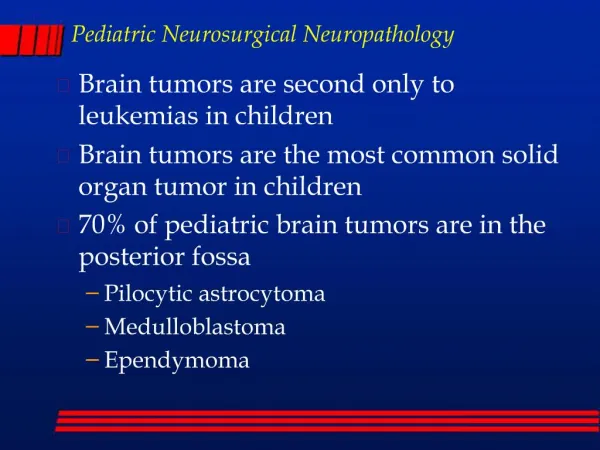
Neoplasia Nervous System • Meningioma • Glial Tumors: Astrocytoma-Glioblastoma; Oligodendroglioma; • Metastatic Lesions
Normal Histology Central Nervous System CORTEX NEURON ASTROCYTE MENINGES OLIGODENDROCYTE
Meningioma • Benign, Adults; Women: Men 3:2 • Multiple lesions associated with NF2 • Arachnoid meningothelial cell • External surfaces: parasagittal convexity • Encapsulated, well defined dural base • Slow growing • Progesterone Receptors
Meningioma DURA
Microscopic • Uniform plump to spindle cells in bundles • Classic “whorling” pattern • Syncytial, transitional, fibroblastic, psammomamatous types • Mitoses, pleomorphism suggest Atypical/Aggressive type
Meningioma Normal meninges Psammomatous meniongioma PSAMMOMA BODY
Meningioma WHORLS Syncytial appearance
Glial TumorsAstrocytoma-Glioblastoma Spectrum • 80% of adult primary brain tumors • Cerebral hemispheres but also brain stem • Deceptively well demarcated grossly • Spreads within brain but no metastasis to other organs • Aggressiveness correlated with tumor grade • WHO grades I-IV
Astrocytoma Grade I-II • Well differentiated fibrillary astrocytoma • Mild to moderate increase in nuclei • Variable nuclear pleomorphism • GFAP positive astrocytic cell processes • Indistinct transition from normal tissue • Tumor cells infiltrate millimeters from main tumor mass
Astrocytoma Grade I-II * * * Mass lesion with infiltration, ill-defined, expanding white matter No hemorrhage or necrosis
Astrocytoma Grade IIIAnaplastic Astrocytoma • Increased dense cellularity • Increased nuclear pleomorphism • Mitoses
Astrocytoma Grade IIIAnaplastic Astrocytoma Hypercellularity Pleomorphism Mitosis
Glioblastoma(Grade IV Astrocytoma)Old name Glioblastoma Multiforme • High cellularity, pleomorphism, mitoses • Necrosis “pseudopalisade) • Vascular Proliferation (Angiogenesis) • Rapid growth, mass lesion effects • Aggressive Lesions < 10% survival in 2 years
Hemorrhagic mass lesion Necrosis Crosses midline Glioblastoma
Glioblastoma PSEUDOPALISADE
Glioblastoma VASCULAR PROLIFERATION NECROSIS
Glioblastoma GFAP Ki67
Glial TumorsOligodendroglioma • 5-15% glial tumors • Cerebral hemispheres, white matter • Well circumscribed, gelatinous, cystic • Sheets of uniform cells with central nuclei and a clear halo of cytoplasm (“fried eggs”) • Calcification, slow growth • Prognostic value loss of 1p and 19q
Oligodendroglioma Non-tumor Tumor Hydrocephalus Diffusely infiltrating oligodendroglioma, expanding corpus callosum and white matter (arrows), and invading the basal ganglion (arrowhead)
Oligodendroglioma “Fried egg” appearance or perinuclear halo
Metastatic Lesions • 25% of intracranial malignant tumors are metastases • Carcinomas most common: lung, breast, kidney • Melanoma • Meningeal carcinomatosis, multiple small nodules of tumor