Download
1 / 77
780 likes | 968 Views
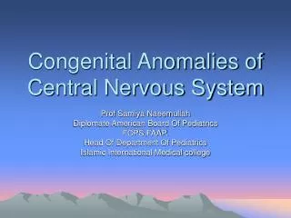
Congenital Anomalies of the Central Nervous System. Major Events in Human Brain Development and Peak Times of Occurrence (Menkes). Gestational Age in Months. Postnatal. 1. 2. 3. 5. 4. 6. 7. 8. 9. Neurulation (3 – 4 wks). Neural tube Brain & Spinal cord Neural crest PNS &

E N D
Major Events in Human Brain Development and Peak Times of Occurrence (Menkes) Gestational Age in Months Postnatal 1 2 3 5 4 6 7 8 9 Neurulation (3 – 4 wks) • Neural tube • Brain & Spinal cord Neural crest • PNS & leptomeninges Prosencephalic (2 – 3 mos) Organization (5 – postnatal years) Neuronal Proliferation (3 – 4 mos) Paired cerebral Hemispheres, LV, BG, Thalami, Optic Nerves/ chiasm CC, SP Myelination (Birth – Postnatal yrs) Synaptogenesis programmed Cell death Full comple- Ment of neu- rons in cerebral hemispheres Neuronal Migration (3 – 5 mos) Formation of myelin electrical conduction Formation from 4 layered embryonic cortex 6 layered adult cortex
Transverse sections of embryos at Different ages to show the Dev’t of the spinal cord
Early development of the human nervous system late presomite and early neural plate stage early somite and neural groove stage eight-somite and early neural tube stage
Congenital Anomalies in the different stages Of CNS development Gestational Age in Months Postnatal 1 2 3 5 4 6 7 8 9 Neurulation (3 – 4 wks) Encephalocoele Myelomenin- gocoele Prosencephalic (2 – 3 mos) Organization (5 – postnatal years) Neuronal Proliferation (3 – 4 mos) Holoprosence- phaly Dandy walker Myelination (Birth – Postnatal yrs) Perinatal Insults ICH/ HIE Tumors CNS Infections Aqueductal Stenosis Hydroceph Perinatal Insults ICH Tumors CNS Infections Neuronal Migration (3 – 5 mos) Schizencephaly Colpocephaly Lissencephaly Agenesis of corpus callosum
Neural Tube Defects (Posterior Midline Defects/Dysraphism) • Results from failure of the neural tube to close spontaneously between the 3rd-4th week of in utero development • Possible etiologic factors: • Radiation • Drugs • Malnutrition • Chemicals • Genetic determinants (mutations in folate-responsive and folate-dependent pathways)
Neural Tube Defects • Spina bifida occulta • Meningocoele/ Myelomeningocoele • Encephalocoele • Anencephaly • Dermal sinus • Tethered cord • Syringomyelia • Diastematomyelia
Neural Tube Defects • Diagnostic tool: • Failure of closure of the neural tube allows excretion of fetal substances (AFP, acetylcholinesterase) into the amniotic fluid • Prenatal screening of maternal serum for AFP during 16-18 week AOG • AF AFP obtained between 15-20 weeks’ gestation is most specific • Rostral end of the NT closes on the 23rd day and the caudal neuropore closes by the 27th day of development
Neural Tube Defects and FA • Maternal periconceptional use of folic acid supplementation reduces the incidence of NT defects by at least 50% • US: recommends all women of childbearing age take 0.4 mg of folic acid daily, and women with previous pregnancy of NT defect should be treated with 4 mg of folic acid beginning one month before pregnancy is planned, until at least the 12th week AOG when neurulation is complete • Fortification of flour, pasta, rice and cornmeal with 0.15 mg of folic acid/100 g was mandated in the US and Canada in 1998
Spina Bifida Occulta • Midline defect of the vertebral bodies without protrusion of the SC or meninges • May be asymptomatic without neuro signs • In some, patches of hair, lipoma, discoloration of skin or dermal sinus may be present
Spina Bifida Occulta • Spine x-ray: defect in closure of the posterior vertebral arches and laminae, usually in L5 and S1 • May be associated with syringomyelia, diastematomyelia, and tethered cord • Recurrent meningitis of occult origin should prompt careful exam for dermal sinus tract
S. N., 2 mos old, female Marked obstructive Hydrocephalus secondary to ARNOLD CHIARI II Neurulation Period Gestational Age in Months Postnatal 1 2 3 5 4 6 7 8 9
Meningocoele • Formed when the meninges herniate through a defect in the posterior vertebral arches • SC may be normal, or may present with tethering, syringomyelia, or diastematomyelia • A fluctuant mass that may transilluminate along the vertebral column
Meningocoele • Dxtic: plain x-ray, utz, MRI for the spine, CT of the head to R/O HCP • Txtic: Asymptomatic children with N neuro findings and full-thickness skin may have surgery delayed. Patients with leaking CSF or a thin skin covering should undergo immediate repair to prevent meningitis.
Meningocoele • Anterior meningocoele may project into the pelvis through a defect in the sacrum causing symptoms of constipation and bladder dysfunction • Female patients may have associated anomalies of the genital tract (rectovaginal fistula, vaginal septa) • Dxtic: plain x-ray, CT, MRI
Myelomeningocoele • Most severe form of dysraphism involving the vertebral column with an incidence of 1/4000 LB • Risk of recurrence after one affected child increases to 3-4% and increases to ~10% with 2 previous abnormal pregnancies • Certain drugs that antagonize folic acid (TMP, AEDs: CBZ, PHY, Pb, primidone) increase the risk of myelomeningocoele • Valproic acid cause NT defects in ~1-2% of pregnancies
Myelomeningocoele • May be located anywhere along the neuraxis but the LS region accounts for 75% of the cases • Extent and degree of the neuro deficit depend on the location • CM: flaccid paralysis, absent DTRs, sensory deficit below the affected level, postural abn of the LE (clubfeet, subluxation of the hips), constant urinary dribbling and a relaxed anal sphincter
Myelomeningocoele • HCP in association with a type II Chiari defect develops in at least 80% with myelomeningocoele • Infants with HCP and Chiari II develop symptoms of hindbrain dysfunction: difficulty feeding, choking, stridor, apnea, VC paralysis, pooling of secretions, spasticity of UEs • Chiari crisis is due to downward herniation of the medulla and cerebellar tonsils
Myelomeningocoele • Requires a multidisciplinary approach: surgeon, therapist, pediatrician • Surgery: repair and shunting; orthopedic procedure, urologic evaluation • GUT: regular catheterization to prevent UTI and reflux leading to PN and hydronephrosis, urine cult, serum elec, creatinine, renal scan, IV pyelogram, Utz • Rehab: functional ambulation (sacral or LS lesion)
Myelomeningocoele • Prognosis: • MR- 10-15% • Most deaths occur before age 4 years • 70% have normal intelligence, but learning problems and seizure disorders are common • History of meningitis or ventriculitis adversely affect the ultimate IQ
11 mos old, male Neurulation Period Gestational Age in Months Postnatal 1 2 3 5 4 6 7 8 9
Encephalocoele • Cranium meningocoele: CSF-filled meningeal sac only • Cranial encephalocoele: contains the sac plus cerebral cortex, cerebellum or portions of the brainstem usually with abnormalities • Usually occurs in the occipital region or below the inion, although in some countries, frontal or nasofrontal encephalocoeles are more prominent
Encephalocoele • Dxtic: • Plain x-ray of the skull and cervical spine • Cranial utz • In utero: AFP, biparietal diameter • Prognosis: • Encephalocoele- at risk for visual problems, microcephaly, MR, seizures • Meckel-Gruber syndrome: AR condition, occipital encephalocoele, cleft lip or palate, microcephaly, microphthalmia, abnormal genitalia, polycystic kidneys, and polydactyly
Anencephaly • Large defect of the calvarium, meninges, and scalp associated with a rudimentary brain which results from failure of closure of the rostral neuropore • Primitive brain consists of portions of connective tissue, vessels and neuroglia • The cerebral and cerebellar hemispheres are usually absent, and only a residue of the brainstem can be identified; the pituitary gland is hypoplastic, and the SC pyramidal tracts are absent
Anencephaly • Associated anomalies: folding of the ears, cleft palate, congenital heart defects (10-20%) • Die within several days of birth • Frequency: 1/1000 LB • Recurrence risk: 4% and increases to 10% with 2 previously affected pregnancies • Monitoring of succeeding pregnancies: amniocentesis, AFP levels, Utz between 14-16th week of AOG
P.M, 22 days old CA 36-37 wks, with cleft lip and palate Holoprosencephaly, semilobar Macrogyria Absent Septum Pellucidum Dysgenesis of the Corpus Callosum
Anterior Midline Defects (Holoprosencephaly) • Alobar, semilobar, lobar, middle interhemispheric fusion variant • Alobar holoprosencephaly: single midline ventricular cavity, absent falx, inferior frontal and temporal regions are absent, only the primary motor and sensory cortex may be present • Failure of cleavage of the hemispheres which occur at 33 days AOG • Defective expression of the gene Sonic hedgehog (Shh) at 7q • Associated with maternal diabetes
Holoprosencephaly • Clinically, present with profound MR, Szs, rigidity, apnea and temperature imbalance; HCP can develop with aqueductal obstruction; endocrine disorders can present with hypothalamic or pituitary malformations • Facial abnormalities include cyclopia, cebocephaly, and premaxillary agenesis • Dxtic: facial x-ray to show deformed anterior craniobasal bones, cytogenetics, MRI. EEG, VER, ABR are generally abnormal.
Disorders of Neuronal Migration • Migrating neurons attach to the radial glial fiber and reach their predetermined sites to form the six-layered cerebral cortex • Small heterotopias- little or no CM • Lissencephaly • Schizencephaly severe MR
Lissencephaly(Agyria) • Absence of the cerebral convolutions and a poorly formed sylvian fissure (3-4 month fetal brain), numerous gray heterotopia • Present with failure to thrive, microcephaly, marked developmental delay, severe seizure disorder, hypoplasia of the optic nerve, microphthalmia • Miller-Dieker syndrome: prominent forehead, bitemporal hollowing, anteverted nostrils, prominent upper lip, micrognathia (90% with chromosomal deletions of 17p13.3- lissencephaly I gene)
Schizencephaly • Unilateral or bilateral clefts within the cerebral hemispheres due to an abnormality of morphogenesis • The cleft may be fused or unfused, and is usually surrounded by abnormal brain, microgyria • Present with severe MR, intractable Szs, microcephaly, spastic quadriplegia when clefts are bilateral
A.M., 2 months old was noted to be microcephalic w/ sz Porencephalic Cyst Schizencephaly Absent Septum Pellucidum Dysgenetic Corpus Callosum Aqueductal Stenosis Neuronal Migration Gestational Age in Months Postnatal 1 2 3 5 4 6 7 8 9
Porencephaly • Cysts or cavities within the brain that may or may not communicate with the ventricular system, resulting from vascular or infectious results during late fetal or early infantile life • Usually present with hemiparesis and focal seizures during the 1st year of life
Agenesis of the Corpus Callosum • Results from an insult to the commissural plate during embryogenesis • When it appears as an isolated phenomenon, the patient may be normal; but those with associated migration defects (heterotopia, microgyria, pachygyria) may present with MR, microcephaly, hemiparesis, diplegia and seizures • CT/MRI: widely separated frontal horns with an abnormally high position of the 3rd ventricle
Agenesis of the Corpus Callosum • Aicardi Syndrome • Patients are almost all females (may be lethal in males) • Characterized by severe MR, intractable seizures with onset between birth and 4 mons of age, and chorioretinal lacunae. Hemivertebrae and costovertebral anomalies are common. • EEG: independent activity from both hemispheres as a result of the absence of the CC
Agenesis of the Cranial Nerves • II, III(Congenital ptosis), V, VIII, IX, X, XI, XII • Marcus Gunn phenomenon: sucking jaw movements causing simultaneous eyelid blinking • Mobius syndrome: absence of the VII nerve resulting in bilateral facial weakness, associated with abducens nerve paralysis. Result in feeding difficulties due to poor suck. Immobile or dull facies may be misinterpreted as MR.
Microcephaly • HC >3 SDs below the mean for age/sex • Primary (genetic) Microcephaly • Secondary (non-genetic) Microcephaly • Dxtic: mother’s serum phenylalanine, karyotype (abnormal facies, short stature, associated congenital abn), CT/MRI (TORCH), fasting plasma and urine amino acid analysis, serum NH4.
Hydrocephalus • CSF production: 20 ml/hr • Total CSF volume: 50 ml (infant) 150 ml (adult) • Normal IV pressure: 180 mm H2O • Communicating Hydrocephalus • Noncommunicating or obstructive hydrocephalus
Hydrocephalus • Obstructive HCP • Aqueductal stenosis- x-linked recessive trait, associated with spina bifida occulta, NF • Aqueductal gliosis- 20 to neonatal meningitis or SAH in PT, intrauterine viral infections, mumps meningoencephalitis • Vein of Galen malformation • Space occupying lesions in the posterior fossa
C. V., 1 month old At 2 weeks of age was noted to have a fast enlarging head Aqueductal Stenosis Neuronal Proliferation Gestational Age in Months Postnatal 1 2 3 5 4 6 7 8 9
Vein of Galen Malformation R.C., 5 months old came in because of enlarge head size
Hydrocephalus • Infant: increasing HC, open and bulging AF, dilated scalp veins, broad forehead, setting-sun eye signs, hyperreflexia, spasticity, clonus, bilateral Babinski sign • Child: irritability, lethargy, poor appetite, vomiting, headache. Gradual personality change and deterioration in academic performance • Serial HC monitoring, Macewen sign • Papilledema
Causes of Hydrocephalus • Chiari malformation • Type I: medulla is displaced caudally into the spinal canal, inferior cerebellar hemispheres is herniated through the foramen magnum • Type II: medulla, cerebellum and part or all of the 4th ventricle are displaced into the spinal canal, with associated obst hcp and LS spina bifida • Type III: features of type I or II plus the entire cerebellum is herniated through the foramen magnum with a cervical spina bifida cystica • Type IV: cerebellar hypoplasia with other malformations of the posterior fossa
Chiari Malformation Type I Type II
Hydrocephalus • Cause: Dandy-Walker malformation- cystic expansion of the 4th ventricle in the posterior fossa • Dxtic: • Plain skull x-ray: separation of sutures, erosion of the post clinoids, beaten-silver appearance • UTZ/CT/MRI • DDx: thickened cranium, metabolic and degenerative disorders producing megalencephaly, gigantism, NF, familial megalencephaly • Management: depends on the cause