Download
1 / 1
10 likes | 109 Views
Study on signaling pathways in human blood monocytes post LPS stimulation, detecting inhibitors of specific pathways. Results show complex interactions post-stimulation.

E N D
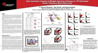
P-S6 Response to LPS Stimulation 0 min 4 min P-p38 Alexa 488 P-S6 Pac Blue P-S6 Pac Blue Ly294002 P-SAPK PE UO126 15 min 60 min 10 min 4 min P-ERK Alexa 647 +UO126 +LPS +UO126 +LPS P-S6 Pac Blue P-S6 Pac Blue + LPS Control P-S6 Pacific Blue P-S6 Pacific Blue P-S6 Pacific Blue +Ly294002 +UO126 +Ly294002 +Ly294002 +UO126 +Ly294002 8 min +UO126 +LPS P-ERK Alexa 488 P-ERK Alexa 488 P-ERK Alexa 488 +Ly294002 +UO126 +Ly294002 Flow Cytometric Analysis of Multiple Signaling Pathways in LPS Stimulated Human Peripheral Blood Monocytes T. Vincent Shankey1, Sue Chow2, and David Hedley21Advanced Technology Group, Beckman Coulter, Inc., Miami, FL, USA2Division of Applied Molecular Oncology, Princess Margaret Hospital, Univ of Toronto, Toronto, Ont., Canada LPS Stimulation of MAP Kinases in Peripheral Blood Monocytes Abstract Background The availability of antibodies specifically reactive with phospho-epitopes characteristic of activated signaling proteins provides specific probes that can be used to simultaneously monitor multiple signal transduction pathways. However, successful use of these probes to intracellular targets has required the development of cell fixation and permeabilization techniques that preserve phospho-epitopes, plus cellular light scatter properties, plus cell surface CD molecules. We have developed specific fixation and permeabilization techniques to monitor signaling pathways in blood and bone marrow samples. We describe an optimized protocol that addresses these issues, and is able to detect inhibitors of specific pathways on multiple signaling targets. Methods Normal whole blood samples are activated with LPS (E. coli from several different strains/purity) at 37 deg C for increasing periods of time, followed by fixation in 4% formaldehyde and subsequent white cell permeabilization and red cell lysis using 0.1% Triton X-100 as previously described (Chow et al Cytometry 2005;67:4-17). Before antibody labeling (surface and cytoplasmic epitopes), cells are treated with 50% MeOH to “unmask” phosphor-epitopes. Whole blood monocytes are labeled using CD14-PECy7. Simultaneous labeling of four independent signaling targets is performed using P-p38T180/Y182–Alexa488 (CST); P-SAPKT183/Y185-PE (CST, custom conjugate by Beckman Coulter); P-ERKT202/Y204–Alexa647 (CST); and P-S6S235/236-Pacific Blue (CST, custom conjugate by Beckman-Coulter). Flow cytometry was performed using a Gallios™ (Beckman Coulter) equipped with violet, blue, and red lasers. Results Normal whole blood Monocytes (CD14+ events) show a complex pattern of signaling responses to LPS stimulation. P-SAPK and P-p38 MAP kinases peak after 10-15 min of LPS stimulation, followed by a decrease in phospho-epitope signals. In contrast, CD14+ cells show a rapid phosphorylation of ERK, peaking at 2-4 min after LPS stimulation, with a descease at 6 min post stimulation, and a subsequent second peak in P-ERK phosphorylation at 10-15 min LPS stimulation. Levels of P-S6 start to increase after 4-6 min stuimulation, with the majority of P-S6+ events also P-ERK+. P-S6 levels remain high for extended periods after activation. Overall, our results show evidence of complex patterns of interactions of signaling pathways after LPS stimulation. Conclusions The LPS stimulation assay outlined here using normal whole blood monocytes provides a method to enable a beginning lab to successfully accomplish flow-based measurements of one or more signaling targets. In addition, it allows testing and validation of reagents for MAPK analysis, including targeted inhibitors that are proposed to selectively target specific targets in specific signaling pathways. Finally, simultaneous analysis of multiple signaling pathways using flow cytometric analysis provides unique insight into the complex biology of signaling in monocytes (using LPS stimulation) and in other blood or bone marrow target cell populations (using appropriate agonists). Both PI3K and ERK Pathways can activate the ribosomal S6 protein Activation of whole blood monocytes in the presence of PI3K and/or ERK Inhibitors Whole blood was activated for the times indicated above with LPS, with or w/o Ly294002 (PI3K inhibitor) and/or U0-126 (MEK inhibitor). Both inhibitors block phosphorylation of S6 at early time points (2-6 min after LPS stimulation). However, at later time points (after 6 min LPS stimulation) the PI3 Kinase inhibitor Ly294002 no longer inhibits phosphorylation of either ERK or S6. The inhibition of P-ERK by a PI3 Kinase inhibitor is unexpected, and implies that early activation of ERK is through the PI3 Kinase pathway. Inhibition of both early (2-3 min) and late (8-15 min) ERK activation by the MEK inhibitor U0-126 indicates that both P-ERK peaks are the result of MEK activation of ERK.. Whole blood was incubated at 37 deg C for 10 minutes with LPS or untreated (Control). Each sample was fixed, permeabilized, and treated with cold 50% MeOH, and washed with PBS/BSA before Incubation with surface (CD14-PECy5) plus antibodies to all 3 MAP Kinases. In bivariate histograms above, CD14+ events are colored in pink. Single parameter histograms show the difference between unstimulated Control sample (red) and sample stimulated with LPS (blue). Materials and Methods LPS (lipopolysaccharide) is known to activate multiple signal transduction pathways in peripheral blood monocytes through its interaction with cell surface Toll-like receptor 4 (TLR4) complex. Following LPS exposure, monocytes rapidly activate three major MAP Kinase pathways – ERK, p38, and SAP/JNK, in addition to activating PI3 Kinase and the IKK/Nf-kappaB pathways. Flow cytometry provides a unique method to simultaneously monitor multiple signaling pathway, allowing coordinated measurements of multiple phospho-epitopes in the context of cell surface plus other cellular markers. Whole blood is obtained from normal donors by veinipuncture, and is collected into K2EDTA vacutainers, and stored at room temperature until used (normally within 3-4 hrs). LPS (100 ng) is added to 100 µl whole blood in 12 X 75 mm polypropylene tubes, mixed by vortex, and the tubes are immediately placed into a 37 deg C bath. For samples containing inhibitors (U0-126 and/or Ly294002), inhibitors are added to whole blood and incubated at 37 deg C for 20 min before addition of LPS. Whole blood fixation, permeabilization and phospho-epitope unmasking are performed as previously described (Cytometry 67A:4-17, 2005). Briefly, LPS activation is stopped by the addition of 65 µl formaldehyde (10%, Polysciences). After addition of formaldehyde, tubes are vortexed and incubated at room temperature for 10 min. 1 ml 0.116% Triton X-100 (Pierce Chemicals) in PBS is added to each tube, vortexed, and incubated at 37 deg C for 10 min. 1 ml cold wash buffer (PBS/4%BSA) is added to each tube, vortexed, and centrifuged at 400 X G for 4 min. After careful removal of all supernate, tubes are vortexed while adding 1 ml cold 50% methanol (in PBS). Tubes are incubated at 4 deg C for 10 min, centrifuged (400 X G for 4 min), decanted, and the pellet is washed with 1 ml cold wash buffer. Following centrifugation (400 X G for 4 min), cell pellets are resuspended in 100 µl antibody cocktail containing optimal concentrations of CD14-PECy7, P-p38(T180/Y182)–Alexa488, P-SAPK(T183/Y185)-PE, P-ERK(T202/Y204)–Alexa647, and P-S6(S235/236)-Pacific Blue, and incubated at room temperature for 30 min. Following centrifugation and resuspension in wash buffer, samples are analyzed on a Gallios™ (Beckman Coulter) flow cytometer equipped with violet, blue, and red lasers. Data is analyzed using CXP ™, or Kaluza ™ (Beckman Coulter), or WinList ™(Verity Software) software. Kinetics of MAP Kinase Activation at 37 deg C LPS Stimulation of MAPK and NF-κB • Conclusions • Simultaneous measurement of multiple signaling epitopes provides important information regarding potential interactions between different pathways. • LPS activation of peripheral blood monocytes stimulates 3 MAP Kinase pathways, plus PI3 Kinase. • Stimulation with LPS at 37 deg C results in two temporally distinct activation peaks for ERK • Our data is consistent with the hypothesis that the first P-ERK peak is a consequence of (early) PI3 Kinase activation that activates the ERK MAP Kinase pathway at some point above MAPKK (MEK). Whole blood was activated at 37 deg C for the times indicated above and analyzed at each time point for the expression of P-ERK, P-p38, P-SAPK (not shown above) and P-S6. Unstimulated peripheral blood monocytes frequently express some P-p38 or P-ERK; as shown above (0 min) simultaneous expression of both phospho-epitopes is rare in unstimulated monocytes. LPS activation results in a rapid activation of P-ERK, and as shown above, at 4 minutes P-S6 expression is seen in cells with the highest levels of P-ERK; all P-ERK+ cells are increasing in the level of expression of P-p38. After 15 min of LPS stimulation, all monocytes show high P-p38, P-ERK, and P-S6 levels, which decrease at later times after LPS stimulation. Of the four signaling markers measured here, P-S6 peaks after all MAP Kinases, and decreases to baseline at a slower rate. The LPS activation of all three MAP Kinases shows a distinct, and reproducible pattern, as shown above. At 37 deg C, LPS activation Of whole blood results in monocytes demonstrating an early peak (~ 2 min) of P-ERK expression, which rapidly decreases, then reverses, and showns a second pear at 10-15 min. This second peak of P-ERK activity correlates with the peak monocyte levels of the other two MAP Kinases, p38 and SAP/JNK P-S6