Download
1 / 19
210 likes | 276 Views
Explore ultrasound elastography for prostate cancer detection. Detailed methods of phantom construction, strain imaging results, and future research directions are discussed.

E N D
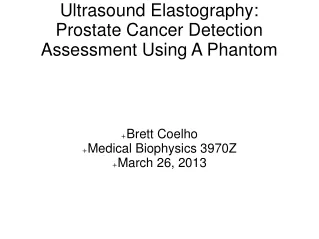
Ultrasound Elastography: Prostate Cancer Detection Assessment Using A Phantom • Brett Coelho • Medical Biophysics 3970Z • March 26, 2013
Introduction • Prevalence and Early Detection • Ultrasound Elastography • Specificity and Sensitivity B-mode Prostate Image
Introduction • Stiffness • Strain Images B- mode Image Strain Image
Methods • Hooke’s Law • Young’s Modulus Reconstruction: Iterative
Methods • Phantom Construction Phantom Ingredients
Methods • Preparing Phantom Solutions • Gelatin – 70 • Agar – 80 • Formaldehyde – 90 • Glycerol – 90 • Sigma Cell • Cool Down
Methods • Pouring The Moulds Prostate and Surrounding Tissue Moulds Tumor Moulds
Methods • Pouring The Moulds Prostate and Sample Moulds
Methods • Uniaxial Test Uniaxial Testing Machine and Cylindrical Sample
Methods • Ultrasound Circular Ultrasound Array Linear Array
Methods • Strain Images Strain Imaging
Results • Uniaxial Results • Ratios Ep/Es=5.4 • Et1/Es=8.7 • Et2/Es = 9.3 Stress kPa Strain
Results • Post Processing • Elastic Moduli Reconstruction Strain Image Frames
Results • Post Processing • Ep/Es=2.2 • Et1/Es=6.0 • Et2/Es=8.9 Phantom Tissue Manual Selections
Results • Tumour Mis Allocations • TM1=5.21 • TM2=5.68 B-mode Image Tumor Locating Reconstructed Image Tumor Locating
Conclusions • Comparing the ratios • Tumor Misallocations
Sources of Error • Strain Images • Uniaxial Test • Assumptions for Stress Calculation
Future Research • Improve Detection and Modulus Calculations • Computational Time • Assumptions and Meshing
Acknowledgements • Dr. Samani • Reza Mousavi • Thank you!