Download
1 / 46
460 likes | 485 Views
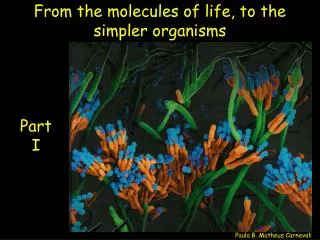
Gain insight into the structure and function of essential biomacromolecules such as carbohydrates, lipids, proteins, and nucleic acids. Explore the formation and breakdown of polymers like starch, cellulose, and triglycerides, and their roles in energy storage and structural support.

E N D
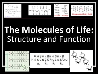
Objective To understand the structure and function of biomac- romolecules and to be able to identify them based on their characteristics. Essential Question: What are the molecules of life, what are their general structures, and functions?
Polymers What is a polymer? Poly = many Mer = part A polymer is a large molecule consisting of many smaller sub-units bonded together. What is a monomer? A monomer is a sub-unit (single unit) of a polymer.
Making and Breaking Polymers How are bonds between monomers formed in the creation of organic polymers? Dehydration Synthesis reactions (“condensation”) Monomers bond to one another through the removal of water.
Hydrolysis Polymers are broken down into monomers. hydro = water lysis = to break/lyse, “loosening” Water is added and the lysis of the polymer occurs.
The 4 classes of Biomacromolecules: Carbohydrates Lipids Proteins Nucleic Acids** = save for later!
Carbohydrates Sugars carbo = carbon hydrate = water Molecular formula (CH2O)n Store energy in chemical structure Glucose: most common monosaccharide produced by photosynthetic autotrophs
Structure of Monosaccharides An OH group is attached to each carbon except one, which is double bonded to an oxygen (carbonyl). Each “carbon” is surrounded by a “hydrate” (water)
Carbohydrates are classified according to the size of their carbon chains, varies from 3 to 7 carbons Triose = 3 carbons Pentose = 5 carbons Hexose = 6 carbons
Structure of Disaccharides Double sugar that consists of 2 monosaccharides, joined by a glycosidic linkage. What reaction forms the glycosidic linkage? dehydration synthesis
Examples of Disaccharides: Lactose = glucose + galactose Sucrose = glucose + fructose
Polysaccharides Structure: Polymers of a few hundred or a few thousand monosaccharides. Functions: energy storage molecules or for structural support
Examples of Carbohydrates Starch = a plant storage from of energy, easily hydrolyzed to glucose units Cellulose = a fiber-like structural material - tough and insoluble - used in plant cell walls Glycogen = a highly branched chain used by animals to store energy in muscles and the liver. Chitin = a polysaccharide used as a structural material in arthropod exoskeleton and fungal cell walls. Lactose = a simple sugar found in milk and dairy products Glucose = simplest sugar; a monosaccharide; feeds brain
Lipids Structure: Greasy or oily nonpolar compounds Functions: Energy storage membrane structure Protecting against desiccation (drying out). Insulating against cold. Absorbing shock Regulating cell activities by hormone actions.
Structure of Fatty Acids Long chains of mostly carbon and hydrogen atoms with a -COOH group at one end. When they are part of lipids, the fatty acids resemble long flexible tails.
Saturated and Unsaturated Fats Unsaturated fats : liquid at room temp one or more -C=C- (double bonds) between carbons in the fatty acids allows for “kinks” in the tails most plant fats Saturated fats: solid at room temp have only single C-C bonds in fatty acid tails all Carbon atoms fully surrounded (“saturated” by H’s) most animal fats
Saturated or Unsaturated? That is the question Saturated fatty acid Unsaturated fatty acid
Structure of Triglycerides 1 glycerol + 3 fatty acids 3 linkages are formed between the –OH group of the glycerol and a –H of the fatty acid. Fatty acids and glycerol bound together by ester bonds.
Phospholipids Structure: 1 glycerol + 2 fatty acids + phosphate group. Function: Main structural component of membranes, where they arrange in bilayers.
Waxes Lipids that serve as coatings for plant parts and as animal coverings.
Steroids Structure: Four carbon rings with no fatty acid tails Functions: Component of animal cell membranes Modified to form sex hormones
Example Lipids • Triglycerides = found in food; energy source • Phospholipids = cell membranes; regulation • Waxes = plant and animal coating; candles • Steroids = components of membranes and for hormones
Proteins Structure: Polypeptide (“many peptide”) chains Consist of peptide bonds between 20 possible amino acid monomers Have a 3 dimensional globular shape
Structure of Amino Acid Monomers Consist of an asymmetric carbon covalently bonded to: Hydrogen Amino group Carboxyl (acid) group Variable R group specific to each amino acid
Properties of Amino Acids Grouped by polarity Variable R groups (side chains) confer different properties to each amino acid: polar, water soluble. non-polar, water insoluble positively charged negatively charged
Structure of Proteins Dehydration synthesis reactions form the peptide bonds between amino acids
Functions of Proteins Enzymes = accelerate specific chemical reactions up to 10 billion times faster than they would spontaneously occur. Structuralmaterials keratin - found in hair and nails collagen - found in connective tissue Contraction actin and myosin fibers that interact in muscle tissue.
Specific binding antibodies that bind specifically to foreign substances to identify them to the body's immune system. Specific carriers membrane transport proteins - move substances across cell membranes blood proteins (hemoglobin), that carry oxygen, iron, and other substances through the body. Signaling hormones such as insulin that regulate sugar levels in blood.
Recap • Carbohydrate Functions: • Examples include: • How? • Lipid Functions: • Examples include: • How? • Protein Functions: • Examples include: • How?
http://www.youtube.com/watch?v=Oz2x_yxPXww&feature=related&safety_mode=true&persist_safety_mode=1&safe=activehttp://www.youtube.com/watch?v=Oz2x_yxPXww&feature=related&safety_mode=true&persist_safety_mode=1&safe=active • http://www.youtube.com/watch?v=lijQ3a8yUYQ&safety_mode=true&persist_safety_mode=1&safe=active
Primary Structure Unique sequence of amino acids in a protein How? Dehydration synthesis between amino acids
Modeling Primary Structure: Salivary Amylase • What do the different colors represent? • How do you think they would interact? • GFCWAQYSSNDCR
Secondary Structure Repeated folding of protein’s polypeptide backbone How? H bonds form between atoms in backbone 2 types: a-helix, b-pleated sheets
Let’s model Secondary Structure • Look at your string of amino acids. • What do the different colors represent? • Note the order of colors. • Take the “backbone” and create some a-helices and some b-pleated sheets.
Tertiary Structure Globular folding! How? Bonding between R groups http://www.youtube.com/watch?NR=1&v=ysPt1lIllcs&safety_mode=true&persist_safety_mode=1&safe=active
Let’s model Tertiary Structure • Note the colors on your polypeptide. • White = hydrophilic • Yellow = hydrophobic • Blue = “negatively charged” • Red = “positively charged” • Green = “sulfur R-group” (bonds only Cysteines)
Quaternary Structure 2 or more polypeptides bonded together How? Attraction between backbones and R groups of neighboring globs
Let’s model Quaternary Structure • Find a neighbor, and attach R groups that might be attracted to each other. What types would?
Good image of protein folding • http://www.umass.edu/molvis/workshop/imgs/protein-structure2.png
Factors That Determine Protein Conformation Depends on physical conditions of environment pH, temperature, salinity, etc. Change in environment may lead to denaturation of protein Denatured protein is biologically inactive Can renature if primary structure is not lost What happens when protein folding goes wrong? http://www.youtube.com/watch?NR=1&v=H2Ouxl_GNjA&safety_mode=true&persist_safety_mode=1&safe=active http://www.youtube.com/watch?v=RNIwwLdDLnI&feature=related&safety_mode=true&persist_safety_mode=1&safe=active
Your Tasks • Protein Activity Wrap Up • Stamps for journal activity (models) • Draw your last diagram! • Enzyme Activity is now extra credit! • “Due” tomorrow for 3 stamps – directions are linked to website • There will be a general enzyme question on the test, so know their functions