Download
1 / 70
740 likes | 1.05k Views
What is a Clinical Laboratory Professional and What Are They Doing With My Body Fluids?. Division of Medical Technology University of Minnesota http://medtech.umn.edu. What is in blood?. Plasma - the liquid portion Platelets - the clot forming components

E N D
What is a Clinical Laboratory Professional and What Are They Doing With My Body Fluids? Division of Medical Technology University of Minnesota http://medtech.umn.edu
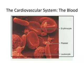
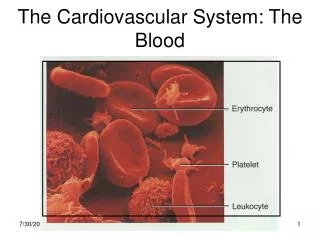
What is in blood? • Plasma - the liquid portion • Platelets - the clot forming components • White Blood Cells - the infection fighters • Red Blood Cells - the oxygen transporters
How do blood tests tell us what’s going on in other parts of the body? • The body of an adult contains over 60,000 miles of blood vessels! • An adult’s heart pumps nearly 4,000 gallons of blood each day! • As the blood travels around the body, products from those cells are absorbed into the bloodstream.
What makes one type of cell different from another type of cell?
CHEMISTRY!! Water Proteins Vitamins Enzymes RNA DNA Cholesterol Amino Acids Minerals Fats Sugars Hormones pH
Chemistry of the Human Body • Each cell of the body contains about 90 trillion atoms (90,000,000,000) • The human body contains about 60 different elements. • Over 96% of those atoms are oxygen (61%), carbon (23%), hydrogen (10%), and nitrogen (2.6%)
How those atoms are put together in the cell determines: • What the cell looks like • What jobs the cell can do in the body • How the cell communicates with other cells in the body
So Bone Cells look and act differently than Blood Cells, Muscle Cells Fat Cells
These chemical differences in cells allow us to determine if something is wrong with the part of the body where that those cells are found. For example …..
What happens in a heart attack? • Blood stops flowing to part of the heart • This can be caused by: • Blood clots • Fat deposits • When blood stops flowing, heart cells don’t get enough oxygen and they begin to die. Blood vessel with fatty deposits
When cells are damaged or dying, they begin to leak some of the compounds from inside their cell membrane. • Those compounds then get into the bloodstream. • Because many of the compounds inside the cell are specific to that type of cell, we can identify problems by the amount of certain compounds in the blood.
What compounds are released by the damaged heart? • Proteins • Myoglobin • Myoglobin is the oxygen-binding protein of the muscles • It only takes 1 to 2 hours after a heart attack before you find myoglobin in the blood • Myoglobin peaks at 6-9 hours. • It returns to normal within 24 hours because it is rapidly excreted in the urine.
What compounds are released by the damaged heart? • Proteins • Troponin-I and Troponin-T • Troponins are structural proteins found in heart muscle. • It only takes 2 to 4 hours after a heart attack before you find troponins in the blood. • Troponin-I peaks at 24 hours. Troponin-T peaks at 12-48 hours. • They remain elevated for 9-14 days.
What compounds are released by the damaged heart? • Enzymes • Creatine Kinase (CK) • It takes 4 to 10 hours before you find the CK enzyme in the blood • It peaks at 24 hours • It returns to normal levels in 2 or 3 days
What compounds are released by the damaged heart? • Creatine Kinase (CK), continued • Different forms of CK, called isoenzymes, are found in different tissues. • CK-MM occurs in high concentrations in skeletal muscle and heart. • CK-MB is present in high concentrations in heart, but it is also present in lungs, small intestine, uterus, prostate, and healthy skeletal muscle. • CK-BB occurs in high concentrations in the brain, but it is also found in lung, stomach, prostate, the gastrointestinal tract, and the bladder.
What compounds are released by the damaged heart? • Creatine Kinase (CK), continued • The different isoenzymes of CK each have a different electrical charge. • This difference in electrical charges allows the lab to separate the CK forms from each other and determine the amount of each form present in the blood by placing the sample in an electrical field. The forms with the greater charge will move the fastest and farthest.
Creatine Kinase Electrophoresis Patterns CK-BB is rarely seen in the blood.
Normal Concentrations CK-MM >95% (Muscle) CK-MB <5 % (Heart) CK-BB 0% (Brain) Normally, there is a small amount of CK-MB in the blood, and almost no CK-BB. After a heart attack, there is more CK-MB in the blood. CK Concentrations in the Blood
What compounds are released by the damaged heart? • Enzymes • Lactate Dehydrogenase (LD) • It takes 6 to 12 hours to appear in the blood • It peaks in 1 to 3 days • Levels return to normal in 8 to 14 days
Determining the amounts of these compounds in the blood helps determine: • Whether or not a heart attack has occurred • How long ago the heart attack occurred • To some extent, how much damage the heart attack caused
In the same way that each type of cell has its own particular chemistry, each disease has its own particular chemistry.
What specimens usually go to the lab for a physical exam? • Two, three, or more tubes of blood • Purple top • Red top • Blue top • Urine specimen
So why do labs need two (or more!) different tubes of blood?Why can’t they just use one tube?
When blood leaves the veins, it will come in contact with factors that will initiate the formation of a blood clot.
The red top tube either contains nothing or something to activate the formation of the clot.The liquid portion is no longer called plasma, but is called serum because it no longer contains the clotting components.Serum is often preferred for testing because all of the cellular components are more easily removed so they can’t clog sensitive instruments.
The purple top tube contains an anticoagulant, EDTA, which prevents the blood from clotting, so cells remain separated. It also preserves the cells so they can be identified by how they look when using particular stains.
The blue top tube also prevents blood from clotting, but it uses a different anticoagulant, sodium citrate, which preserves more of the clotting factors.This is the tube used to evaluate the ability of the blood to clot.
Specimen Processing • The red top tube is centrifuged to separate the serum from the cells. • The blue top tube is also centrifuged and then sent to the coagulation laboratory. • The purple top tube is not centrifuged, but sent to hematology so they can begin to prepare slides and look at the cells. Centrifuge
The serum can now be tested for: • Cholesterol • Blood glucose (sugar) • Electrolytes (sodium, potassium, chloride) • Enzymes from the heart, liver, pancreas, etc. • Kidney function (blood urea nitrogen or creatinine) • Hormones • Vitamins • Drug levels • Hundreds of other compounds
The Purple Top Tube Goes to the Hematology Lab • This tube is mixed • well, then placed • in an instrument • that determines: • Red blood cells • • White blood cells • • Platelets • • Hematocrit (percentage of blood that is cells) • • Hemoglobin (oxygen-carrying protein)
Blood smears are made onglass slides and then stained so cell structures can be seen clearly.
Red Blood Cell Size and Color are Studied NORMAL RED BLOOD CELLS Normal area of central pallor should be about one-third of the cell’s diameter. HYPOCHROMIC Decreased hemoglobin concentration in pale cells. ANISOCHROMIA Pale and filled cells can both be seen following blood transfusion.
Red Blood Cell Shape is Observed Target Cells - Abnormal hemoglobins, liver disease, thalassemias Elliptocytes - Hereditary condition, some anemias Sickle Cells - Sickle cell anemia (Hb SS), Hb SC. Hb S/thal
More Red Blood Cell Shapes Schistocytes - RBC fragments found in severe burn patients, disseminated intravascular coagulation Spherocytes - Red cells without a pale center are seen in hereditary spherocytosis, some immune-mediated hemolytic anemias Acanthocytes - “Spiked” red cells are observed in liver disease, asplenic patients (spleens removed)
White Blood Cells Neutrophils - Engulf bacteria and cellular debris 37-77% Basophils - Hypersensitivity, Release histamine 0-1.6% Eosinophils - Parasitic infections, Allergic response 1-7% Monocytes - Engulf cellular debris, antigen processing 2-10% Lymphocytes - Produce antibodies, regulate the immune response 10-44%
Abnormal Blood Smears Chronic Myelogenous Leukemia Infectious Mononucleosis (Reactive Lymphocytes) Malaria (Plasmodium falciparum)
The Blue Top Tube Goes to the Coagulation Lab • These tubes are often drawn on people who are on anti-coagulant therapy (coumadin), also called “blood thinners” The plasma is tested to determine how long it takes blood to clot.
The Urine Goes to the Urinalysis Lab Urine can provide information about: • Kidney function • Urinary tract disorders • Diabetes • Liver disorders • Metabolic disorders • Muscle trauma • Hormonal disorders • Drug use
Urinalysis A complete urinalysis consists of examining the physical, chemical, and microscopic characteristics of the urine.
Chemical analysis is performed using a reagent strip with pads containing chemicals specific for each test.
What chemicals do labs test for in urine? • Sugar • Normal urine should not contain glucose (blood sugar) • Found in diabetes • Ketones • Normal urine should not have ketones • Ketones form when you break down fats instead of sugar for energy • Found in diabetes, starvation
What chemicals do labs test for in urine? • Protein • Normal urine should contain very little protein • Suggests possible kidney damage, certain cancers • Hemoglobin • Normal urine should not contain hemoglobin (from red blood cells) • Seen in injury, kidney stones, infection
What chemicals do labs test for in urine? • Leukocyte esterase • Normal urine should not contain this enzyme found in white blood cells • Suggests urinary tract infection • Nitrite • Normal urine should not contain nitrites • Nitrites form when bacteria convert urine nitrates to nitrite
Urine MicroscopicExam Yeast Epithelial Cells Calcium Oxalate Crystals
Clinical Microbiology Identify organisms Determine antibiotic sensitivity
The virology lab tests the serum for antibodies directed against viruses such as HIV or hepatitis.