Download
1 / 1
10 likes | 142 Views
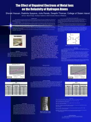
The Effect of Unpaired Electrons of Metal Ions on the Relaxivity of Hydrogen Atoms. Shorok Hassan, Radmila Ilyayeva, Julia Rando, Deepthi Thomas: College of Staten Island. Advisor: Sheref Girgis, Graduate Student at Albert Einstein School of Medicine.

E N D
The Effect of Unpaired Electrons of Metal Ions on the Relaxivity of Hydrogen Atoms Shorok Hassan, Radmila Ilyayeva, Julia Rando, Deepthi Thomas: College of Staten Island Advisor: Sheref Girgis, Graduate Student at Albert Einstein School of Medicine What is Magnetic Resonance Imaging (MRI)? Magnetic resonance imaging is a technique used to describe an image of the internal structure, or anatomy, of a patient. The ideal atom for MRI is the hydrogen atom, because its nucleus has a single proton and a large magnetic moment. The basics of a MRI include the use of an electromagnet and radio waves. The electromagnet aligns the hydrogen atoms in the body magnetically. Different forms of transition elements, such as Fe, are used as electromagnets because the 3d sublevel of electrons is partially empty. This results in more unpaired electrons. When a patient is lying on his or her back in the MRI scanner, the hydrogen protons in his or her body will line up in the direction of either the feet or the head. A great amount of these protons will cancel each other out. This is because for each hydrogen proton lined up toward the feet, one toward the head will cancel it out. Only a few protons out of every million are not canceled out, and these “extra” protons are responsible for producing the image. In the next step, the MRI machine applies a radio frequency (RF) pulse, specific only to hydrogen. The pulse, or radio waves, is directed toward the part of body being analyzed. The pulse causes the protons in that area to absorb enough energy so that they precess, or spin, in a different direction. Thus, they are flipped. When these pulses are removed, the hydrogen protons relax back to their original positions within the magnetic field. As they relax, excess stored energy is released. This produces a signal that a coil picks up and sends to the computer system, forming a picture. INTRODUCTION This experiment was performed to determine the effect of unpaired electrons of metal ions on the relaxivity of hydrogen atoms. Relaxation is the measurement of how fast these hydrogen atoms relax, while relaxivity is a measurement of the ability of a magnet (Fe) to make the atoms relax. Fe (II) has four unpaired electrons. Fe (III) has five unpaired electrons. It was hypothesized that Fe (III) would cause the hydrogen atoms to relax faster because it has more unpaired electrons. A ligand is a molecule or ion that is bonded directly (i.e. covalently) to a metal center. This results in an inability of the electrons to get close to the hydrogen atoms. In this experiment the ligand, ethylenediaminetetraacetate (EDTA), was used to further prove that it is the unpaired electrons of the transition metal that cause the relaxation of hydrogen atoms. According to the hypothesis, when the Fe is wrapped in EDTA, it should take longer for the hydrogen atoms to relax; therefore, the negatively charged electrons of Fe will be unable to get close to the positively charged hydrogen atoms. This experiment can be applied to magnetic resonance imaging (MRI). The MRI technology has allowed this technique of MRI to be used extensively in New York. The science and technology related to MRI has become extremely helpful in treatment planning and also post treatment follow-up for many types of diseases. By applying the research of this experiment, the effect of the unpaired electrons of the electromagnetic core on relaxation of hydrogen atoms can be determined. The hypothesis, thus, is the more unpaired electrons and a shorter distance from the hydrogen atoms will result in a faster relaxation time of the atoms, and therefore, a clearer image. ABSTRACT In order to determine the effect that unpaired electrons of metal ions have on the relaxivity of hydrogen atoms., six different concentrations of Fe (II) (aq) and Fe (III) (aq) were tested by a minispec, which calculated the relaxation times of the hydrogen atoms. It was hypothesized that the metal ion with more unpaired electrons, Fe (III), would cause the hydrogen atoms to relax more quickly (Volkov 1997). A second trial was then performed, in which 223.44mg of ethylenediaminetetraacetate (EDTA) was added as a ligand to each concentration of Fe (II) (aq) and Fe (III) (aq), and these samples were also run through the Minispec. The relaxation times of the hydrogen atoms from both trials was recorded in Table I and II. This data was then plotted as 1/relaxation time v. concentration in order to determine the relaxivity of each solution, in Figure I and II. From these plots, it was determined that unpaired electrons do cause hydrogen atoms to relax more quickly. This research can be applied to Magnetic Resonance Imaging (MRI) because a clearer image will be produced as result of a faster relaxation time of the hydrogen protons. EDTA (HOOCCH2)2NCH2CH2N(CH2COOH)2 • PROCEDURE • Each metal is tested in 6 different concentrations: 6M, 5M, 4M, 3M, 2M, and 1M. In order to produce the 6M solution, a specific amount of the metal is dissolved in water in a 100ml volumetric flask. This solution is used to produce the other concentrations. • Without EDTA Ligand: • The first metal tested is Fe (II). In order to produce a 6M solution of Fe (II), 166.81 mg were measured and dissolved in a 100ml volumetric flask. Using a pipette, a NMR tube is filled with about 1cm of the 6M solution. The tube is placed in the minispec, and a relaxation time (T1) is recorded as it appeared on the computer screen (Table 1). This was repeated with a 5M, 4M, 3M, 2M, and 1M solution of Fe (II), and each • T1 was recorded. The second metal tested is Fe (III). In order to produce a 6M solution of Fe (III), 242.44 mg were measured and dissolved in a 100ml volumetric flask. Using a pipette, a NMR tube is filled with about 1cm of the 6M solution. The tube is placed in the Minispec, and a relaxation time (T1) is recorded as it appeared on the computer screen (Table 2). This was repeated with a 5M, 4M, 3M, 2M, and 1M solution of • Fe (III), and each T1 was recorded. • With EDTA Ligand: • For the second half of the experiment, this entire procedure was repeated. However, 223.44mg of EDTA was added to each of the original solutions (6M of Fe (II) and 6M of Fe (III)). Each relaxation time was recorded • in Table I and Table II. A plot of 1/Relaxation Time Vs Concentration was graphed for each test. MATERIALS The materials used in this experiment include Fe (II), Fe (III), ethylenediaminetetraacetate (EDTA), two 100ml volumetric flasks and stoppers, weigh boats, spatulas, scale, pipettes, water, measuring cylinders, nuclear magnetic resonance tubes (NMR) tubes, a Minispec, and a computer. The Minispec is used to measure the relaxation time of atoms, which is then graphed onto the computer. DATA and ANALYSIS The data gathered from the minispec was recorded in Tables I and II. This data was then plotted as 1/relaxation time v. concentration in order to determine the relaxivity of each solution, in Figure I and II. The slope of the trend lines in each figure is proportional to the relaxivity of the solution times a constant. The slopes of free Fe (II) (m = 4.0 * 10-4) and free Fe (III) (m = 6.7 * 10-3) indicate that Fe (III), with its greater number of unpaired electrons, caused the hydrogen atoms to relax more quickly. In addition, Figure II also supports the hypothesis, as free Fe (III) has a greater relaxivity than the wrapped Fe (III) (m = 2.1 * 10-3). However, in Figure I, a different trend is seen; free Fe (II) has a lower relaxivity than wrapped Fe (II) (m = 1.3 * 10-3). It is believed that there may be an error in this data set because the R2 value = 0.8992, which indicates the data points are not close to a straight line, as compared to the other sets that are much closer to a straight line, where R2 = 0.9901 (free Fe (II)), 0.9957 (free Fe (III)), and 0.9536 (wrapped Fe III). As a result, the EDTA trials for Fe (II) indicate that potential errors may have occurred and a new trial may be conducted in the future. Figure I: Graph of Free and Wrapped Fe (II) Figure I: Graph of Free and Wrapped Fe(III) POTENTIAL ERRORS There are possible errors that could have occurred. One possible explanation for the unexpected results for the wrapped Fe (II) it that the EDTA used to wrap the metal could have been aged. This old EDTA may not have produced the expected reaction. The solutions of Fe and EDTA should have been red. However, the solution containing the Fe (II) and EDTA was a cloudy yellow. The cloudy yellow color of the wrapped Fe (II) solution may have been the result of the metal being oxidized (lost electrons), as can be seen in rust. This oxidation reaction could have prevented the proper interaction between the Fe (II) and the EDTA, producing an erroneous graph of the relaxation times given by the Minispec computer. REFERENCES Hornak, Joseph P. The Basics Of MRI . 1996-2004. 25 Oct. 2004. <http://www.cis.rit.edu/htbooks/mri/inside.htm>. Zeman, Gary. “Magnetic Resonance Imaging.” Health Physics Society. 1 Jan. 2004. 25 Oct. 2004. <http://www.hps.org/hpspublications/articles/mri.html>. Volkov, Andrei. “Contrast Agentsin Magnetic Resonance Imaging.” University of Utah. 23 May. 1997. <http://www.cc.utah.edu/~av6a51/mri.htm>. Moulé , Adam J. “Amplification of xenon NMR and MRI by remote detection.” Proceedings of the National Academy of Sciences of the USA. Vol. 100. No. 16. p. 9122-9127. 5 Aug. 2003. 25 Oct. 2004. <http://www.pnas.org> Sinex, Scott A. “EDTA - A Molecule with a Complex Story.” Prince George's Community College. Mar. 2004. 1 Nov. 2004 <http://www.tlchm.bris.ac.uk/motm/edta/edtah.htm>. Ronen, Itamar. “Imaging of H217O distribution in the brain of a live rat by using proton-detected 17O MRI.” Proceedings of the National Academy of Sciences of the USA Vol. 95. No. 22. p. 12934-12939. 27 Oct. 1998. 1 Nov. 2004. <http://www.pnas.org> CONCLUSION The proposed hypothesis was confirmed. The metal with the most unpaired electrons (Fe III) showed the best results. The slope of free Fe(III) was larger than the slope of free Fe (II), meaning Fe (III) caused the hydrogen atoms to relax faster. Thus, it had the fastest relaxivity, and would, therefore, result in a clearer picture. The hypothesis was further confirmed because the EDTA wrapped Fe (III) showed a smaller slope, slowing down the relaxation time of the hydrogen atoms. This shows that the number of unpaired electrons and their distance from the hydrogen atoms affects the relaxation of the atoms. Therefore, the metal with more unpaired electrons and a shorter distance from the hydrogen atoms will result in a faster relaxation time of the atoms. In relation to MRI, a faster relaxation time will result in a better, clearer picture, since magnet strength equates with better image quality and better patient care. Magnetic Resonance Image of the lumbar spine (normal) ACKNOWLEDGEMENTS Thanks and appreciation to Sheref Girgis (Albert Einstein Medical Student), the Albert Einstein School of Medicine for use of laboratory, and Professor Charles Liu at the College of Staten Island.