Download
1 / 42
420 likes | 623 Views
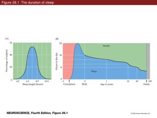
Figure 28.1 The duration of sleep. Figure 28.1 The duration of sleep (Part 1). Figure 28.1 The duration of sleep (Part 2). Figure 28.2 Circadian rhythmicity of core body temperature and growth hormone & cortisol levels. Box 28A Sleep Styles in Different Species.

E N D
Figure 28.2 Circadian rhythmicity of core body temperature and growth hormone & cortisol levels
Figure 28.3 Consequences of total sleep deprivation in rats (Part 1)
Figure 28.3 Consequences of total sleep deprivation in rats (Part 2)
Figure 28.4 Rhythm of waking and sleeping in isolation, with and without day–night cycle cues
Figure 28.5 Photoreceptors responsible for signaling circadian light changes
Figure 28.5 Photoreceptors responsible for signaling circadian light changes (Part 1)
Figure 28.5 Photoreceptors responsible for signaling circadian light changes (Part 2)
Figure 28.5 Photoreceptors responsible for signaling circadian light changes (Part 3)
Figure 28.7 Physiological changes during the various sleep states
Figure 28.7 Physiological changes during the various sleep states (Part 1)
Figure 28.7 Physiological changes during the various sleep states (Part 2)
Figure 28.7 Physiological changes during the various sleep states (Part 3)
Figure 28.8 Circuitry involved in decreased sensation and muscle paralysis during REM sleep
Figure 28.8 Circuitry involved in decreased sensation and muscle paralysis during REM sleep
Figure 28.9 Activation of specific neural circuits triggers sleep and wakefulness
Figure 28.10 Cortical regions whose activity changes during REM sleep
Figure 28.11 Important nuclei in regulation of the sleep–wake cycle
Figure 28.11 Important nuclei in regulation of the sleep–wake cycle (Part 1)
Figure 28.11 Important nuclei in regulation of the sleep–wake cycle (Part 2)
Figure 28.11 Important nuclei in regulation of the sleep–wake cycle (Part 3)
Figure 28.11 Important nuclei in regulation of the sleep–wake cycle (Part 4)
Figure 28.12 Thalamocortical neuron activity in sleep and awake states
Figure 28.13 Thalamocortical feedback loop and the generation of sleep spindles
Figure 28.13 Thalamocortical feedback loop and the generation of sleep spindles (Part 1)
Figure 28.13 Thalamocortical feedback loop and the generation of sleep spindles (Part 2)
Figure 28.15 Sleep pattern of a patient with obstructive sleep apnea