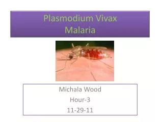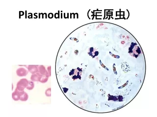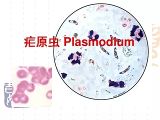
PLASMODIUM
PLASMODIUM. II MBBS Dr Ekta Chourasia Microbiology. Taxonomy. Phylum Apicomplexa Subphylum Sporozoa Genus Plasmodium Disease Malaria

PLASMODIUM
E N D
Presentation Transcript
PLASMODIUM II MBBS Dr Ekta Chourasia Microbiology
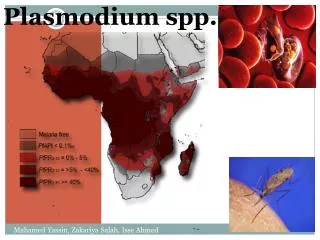
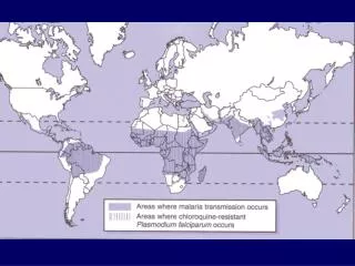
Taxonomy Phylum Apicomplexa Subphylum Sporozoa Genus Plasmodium Disease Malaria Geographical Tropical & distribution Subtropical countries Dr Ekta Chourasia, Microbiology
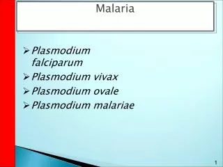
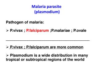
Genus Plasmodium • Consists of 4 species: • P. vivax • P. falciparum • P. malariae • P. ovale Dr Ekta Chourasia, Microbiology
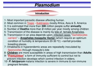
Landmarks in the evolution of Malaria • 1880 – Laveran identified the malarial parasite in an unstained smear • 1885 – Golgi described the blood stage (erythrocytic schizogony) of malarial parasite – Golgi cycle • 1898 – Amigo & Grassi described the life cycle • 1891 – Romanowsky introduced the staining method • 1897 – Ronald Ross while in Calcutta, India, demonstrated Anopheles sp. of mosquitoes as vectors of malaria. Got Nobel prize for his work in 1902 Dr Ekta Chourasia, Microbiology
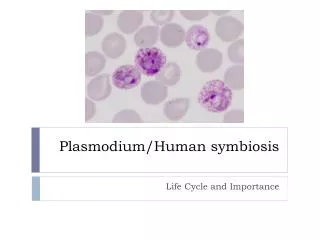
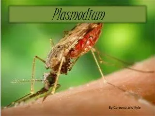
Transmission & Life Cycle Definitive host Female Anopheles mosquito Intermediate host Man Infective form Sporozoites Portal of entry Skin Mode of transmission Bite of an infected mosquito Site of localization First in liver cells & then in RBCs Dr Ekta Chourasia, Microbiology
Phases of Development in Man 2 phases of development • Inside the liver (tissue phase) • Pre- erythrocytic schizogony – no clinical symptoms, no pathological damage • Exo- erythrocytic schizogony – cause of relapse • Inside the RBCs (erythrocytic phase) • Erythrocytic schizogony – cause of malarial paroxsyms • Gametogony – infects mosquito Dr Ekta Chourasia, Microbiology
Morphological forms seen in Humans • In liver: • Sporozoites • Pre erythrocytic schizonts • Merozoites – infect RBCs • In RBCs : • Trophozoites – ring form • Schizonts • Merozoites – released by the rupture of schizonts – infect other RBCs • Gametocytes – micro and macro gametocytes Dr Ekta Chourasia, Microbiology
Morphological forms seen in Mosquito • Further differentiation & development of gametocytes take place in mosquito • Macro gametes (female gametes) – each macro gametocyte develops in to one macro gamete in the mid gut of mosquito • Micro gametes (male gametes) – one micro gametocyte produces 6 to 8 micro gametes by exflagellation. • Zygote– Ookinete – Oocyst – rupture – release of Sporozoites – predilection to salivary glands. Dr Ekta Chourasia, Microbiology
Other modes of transmission • Sporozoite- induced- malaria : injection of an emulsion of salivary glands of mosquito containing sporozoites • Trophozoite- induced- malaria : injection of blood from a malarial patient containing the asexual forms of erythrocytic schizogony e.g. • Transfusion malaria – when persons with latent infection are used as donors • Congenital malaria – transmission through some placental defects (a healthy placenta acts as a physiological barrier) • Drug addicts – by using same syringe Dr Ekta Chourasia, Microbiology
Incubation period • P. vivax • P. ovale 10 to 14 days • P. falciparum • P. malariae 18 days to 6 weeks Dr Ekta Chourasia, Microbiology
Pathogenicity • Infection causes intermittent fever – Malaria • Each of the 4 species causes a characteristic fever: P. vivax Benign tertian/ vivax malaria P. falciparum Malignant tertian/ falciparum malaria, black water fever P. malariaeQuartan malaria P. ovaleTertian/ Ovale malaria Dr Ekta Chourasia, Microbiology
Clinical Features • Series of febrile paroxysms – fever is caused by the release of merozoites & toxins from ruptured erythrocytic schizont which in turn causes the release of cytokines. Quartan malaria – every 72 hrs Tertian malaria - every 48 hrs * each paroxysm has 3 stages - cold stage (rigors), hot stage (high temp., body & joint pains, vomiting & diarrhoea) and perspirationstage (fall in temp.) Dr Ekta Chourasia, Microbiology
Clinical Features • Anaemia – due to breakdown of RBCs, particularly occurs in falciparum malaria • Splenomegaly – all forms Dr Ekta Chourasia, Microbiology
Falciparum Malaria • Most widespread • Accounts for 80% of malaria cases worldwide • Most pathogenic of human malaria species • Untreated infections - severe disease & even death, particularly in young children, pregnant woman & non immune adults. Dr Ekta Chourasia, Microbiology
Falciparum malaria • Severe falciparum malaria is associated with • Pernicious malaria /cerebral malaria • Blackwater fever • Anaemia • Hypoglycaemia • Hypotension • Complications in pregnancy Dr Ekta Chourasia, Microbiology
Pernicious Malaria • Def: refers to a series of phenomenon occurring during infection with P. falciparum which, if not effectively treated, threatens the life of the patient with in 1 to 3 days • In children & non immune adults, can cause coma & death – Cerebral malaria. • Occurs as a result of capillary blockage. Dr Ekta Chourasia, Microbiology
Black Water Fever • Occurs in previously infected subjects • Can also occur in non immune adults with severe falciparum malaria, and also as a complication of quinine therapy. • A rare but acute condition characterised by sudden & massive hemolysis of parasitised & non parasitised RBCs followed by fever and haemoglobinuria. • Often fatal due to renal failure Dr Ekta Chourasia, Microbiology
Black Water Fever • Difficult to find the parasites in the blood following a hemolytic attack. • Urine appears dark red to brown black due to the presence of free Hb. • Clinical features – fever, rigor, aching pains in the loin, icterus, bilious vomiting, circulatory collapse, haemoglobinuria & acute renal failure. • Treatment – Chloroquine, blood transfusion, peritoneal dialysis in ARF. Dr Ekta Chourasia, Microbiology
Anaemia • Can be severe & occur rapidly, particularly in young children • Occurs due to destruction of parasitisedRBCs – phagocytosis & destruction in the spleen • Decreased production of RBCs in the bone marrow. Dr Ekta Chourasia, Microbiology
Falciparum malaria in Pregnancy Can result in: • Severe anemia • Low birth weight babies • Greatest risk in 1st pregnancy Dr Ekta Chourasia, Microbiology
Malaria caused by P.vivax, P.ovale & P.malariae • Rarely life threatening • Relapses/ recurrences are a feature Recurrences in Malaria • May result from – reinfection or - due to certain events related to the parasite’s life cycle Dr Ekta Chourasia, Microbiology
Recurrence of Malaria • Two types of recurrences known in malaria: • Recrudescence – • seen in P. falciparum & P. malariae • due to persistence of blood infection (some erythrocytic forms evade host immunity) even after clinical illness has subsided. • The numbers may increase later, leading to reappearance of clinical symptoms • Occur mostly up to one year or so but in P. malariae, it can occur even after decades Dr Ekta Chourasia, Microbiology
Recurrence of Malaria • Relapse • Occurs due to a special form of parasites – hypnozoites. • Hypnozoites are the sporozoites that remain dormant after infecting liver • Activated from time to time to initiate pre erythrocytic schizogony - Exoerythrocytic schizogony Dr Ekta Chourasia, Microbiology
Genetic factors protecting against Malaria • Sickle cell anaemia – sickle celled RBCs are removed by the spleen before the development of schizonts • Ovalocytosis – RBCs are rigid and they resist parasitic invasion • Duffy blood group negative individuals – duffy blood group Ag is the receptor for the attachment of merozoites of P.vivax Dr Ekta Chourasia, Microbiology
Genetic factors protecting against Malaria • Newborn infants – natural protection for 1st few months of life due to high conc. of HbF in their RBCs. • Beta thalassaemia – protects against severe falciparum infection Dr Ekta Chourasia, Microbiology
Laboratory Diagnosis • Microscopy – detecting & identifying malarial parasites in peripheral blood films. • Concentrating parasites in venous blood by centrifugation when they can not be found in blood films • Using a rapid malaria Ag or enzyme detection test • Other tests – Hb, PCV, Blood glucose, total WBC & platelet count. Dr Ekta Chourasia, Microbiology
Examination of Blood film • Collection of blood - best prepared directly from capillary blood - in EDTA bulb (used within 30 mins) • Time of collection - as soon as possible if malaria is suspected - before administering antimalarials - during pyrexial phase Dr Ekta Chourasia, Microbiology
Types of Blood film • Two types: • Thick films : - 30 to 40 times more sensitive than thin films - more suitable for detection of malarialparasitewhen they are few in number - blood is not fixed, RBCs are lysed during staining (only parasitic forms will be seen) Dr Ekta Chourasia, Microbiology
Types of Blood film 2. Thin films : - to confirm the Plasmodium species - assists in the identification of mixed infections - blood is fixed, parasites are seen within the RBCs - also helps in assessing the response totreatment especially in areas where drug resistance is suspected (by counting the number of parasitised RBCs before & after the treatment) Dr Ekta Chourasia, Microbiology
Making of Thin & Thick films Dr Ekta Chourasia, Microbiology
Fixation & Staining • Fixation – thin films are fixed with absolute alcohol for 1 to 2 mins. • Staining – films are stained with Romanowsky stain: giemsa, field’s, wright’s • Giemsa – 10% solution for 10 mins Dr Ekta Chourasia, Microbiology
Reporting of Blood film • Look for the different morphologicalforms of parasite in blood smear: • Trophozoites / ring forms • Schizont • Gametocytes • Identify species – differences in the characteristics of morphological forms in different species Dr Ekta Chourasia, Microbiology
Trophozoites / Ring forms Dr Ekta Chourasia, Microbiology
Thin Blood Film Thick Blood Film Ring Forms / Trophozoites Dr Ekta Chourasia, Microbiology
Schizont Dr Ekta Chourasia, Microbiology
P. vivax P.falciparum Dr Ekta Chourasia, Microbiology
Gametocytes (male & female) Dr Ekta Chourasia, Microbiology
P.vivax P. falciparum Dr Ekta Chourasia, Microbiology
Counting the % age of parasitised RBCs • On thin blood films • When falciparum malaria parasitemia is high • Method of counting: • Select an area where no of RBCs is roughly 250. • Count the no of parasitised RBCs in 4 such fields i.e. approximately 1000 RBCs. • Divide by 10 to obtain the percentage. *WHO – if it is >5%, then the parasitemia is heavy & prognosis is poor. Dr Ekta Chourasia, Microbiology
Buffy Coat preparation • To concentrate malarial parasite • Centrifuge EDTA anticoagulated venous blood in a thin bore capillary tube • Buffy coat layer is formed between the RBCs & the plasma. • Break the tube & transfer buffy coat & RBCs to a slide - make a thin smear – air dry – fix with ethanol – stain with Giemsa. Dr Ekta Chourasia, Microbiology
Quantitative Buffy Coat • Capillary tube is coated with an anticoagulant & Acridine orange fluorescent dye • After centrifugation, the tube can be used for two purpose: • Complete blood count • Identification of malarial parasite using a fluorescence microscope. Dr Ekta Chourasia, Microbiology
Quantitative Buffy Coat Dr Ekta Chourasia, Microbiology
Rapid Diagnostic tests • Developed to diagnose falciparum malaria rapidly & without a microscope. • Can also detect vivax malaria • Three tests are available commercially • Detects either HRP2 Ag (Histidine rich protein) or specific pLDH (parasite lactate dehydrogenase) • Both HRP2 & pLDH are produced by the parasites during their growth & differentiation in RBCs. Dr Ekta Chourasia, Microbiology
Rapid Diagnostic tests HRP2 tests • detection of P.falciparum • Two types of test – ParaSight F - ICT Malaria Pf pLDH test e.g. OptiMAL test • Detection of P.falciparum & P.vivax • Produced by all human malarial parasites • Differentiation of species is based on antigenic differences between pLDH isoforms. Dr Ekta Chourasia, Microbiology
ParaSightF test Optimal test ICT Malaria Pf / Pv Dr Ekta Chourasia, Microbiology
Stage specificity of antimalarial drugs Dr Ekta Chourasia, Microbiology