The Role of Spectrin Repeat Domains in Cell Function and Associated Genetic Diseases
200 likes | 325 Views
Spectrin repeat domains, composed of three-helix bundles, are crucial for maintaining cytoskeletal integrity and facilitating protein interactions. These repeats can be found in various proteins, including spectrin, dystrophin, and α-actinin, and are integral to cellular structure and function. Recent studies have also identified their significance in disease, linking mutations in spectrin repeat genes to conditions like Duchenne muscular dystrophy (DMD), spinocerebellar ataxia type 5 (SCA5), and hereditary spherocytosis. Understanding these domains opens avenues for targeted therapies.

The Role of Spectrin Repeat Domains in Cell Function and Associated Genetic Diseases
E N D
Presentation Transcript
Spectrin Louise Williams 21st February 2008
Spectrin repeat domains • Spectrin repeats are three-helix bundle structures which occur in a diverse range of proteins as single copies or as multiple tandem repeats. • Spectrin repeats can have structural functions (e.g. co-ordination of cytoskeletal interactions with high spatial precision) or as a switchboard for interactions with proteins in a regulatory role.
Structure • The spectrin repeat domain is composed of three α-helicies. • Aromatic residues in the hydrophobic core of the structure are conserved. • Spectrin helicies have a characteristic length and left-handed twist (due to the gentle curve of the helicies). • Typically, there are between 4 and 20 consecutive repeats in genes.
Function 1 • Traditionally, spectrin repeats have been viewed as ‘spacer’ molecules. • This is because they normally occur in multiple copies and seem to separate other functional parts of proteins. • For example, in the actin cross-linking proteins of the spectrin super family, they control the specific distance between N- and C- terminal functional domains.
Function 2 • Spectrin repeats have recently been discovered to also act as a docking surface for cytoskeletal and signal transduction proteins. • Spectrin repeats have also been found to have elastic properties that might be important for the deformability of the cell cortex.
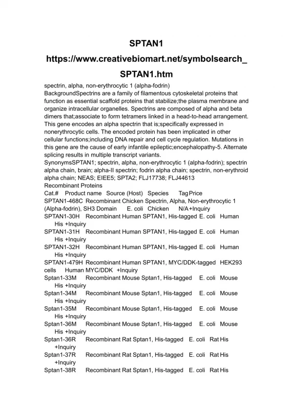
Genes with spectrin repeats • Spectrin repeats can be found in numerous genes. • The best known are the spectrin super family including spectrin, α-actinin, dystrophin and utrophin. • Other proteins have been identified with spectrin repeats without the other characteristic features of this super family.
Associated diseases • There are numerous diseases associated with genes that include spectrin repeat domains. • Some examples are: • DMD associated with dystrophin • SCA5 associated with βIII spectrin • Hereditary spherocytosis and elliptocytosis.
DMD 1 • Duchene muscular dystrophy (DMD) is a rapidly progressive muscle wasting disorder. • DMD is caused by mutations in the dystrophin gene, which is located at Xp21.2.
DMD 2 • The dystrophin protein contains an N-terminal domain that binds to actin, a 24 repeat spectrin domain, a cysteine-rich calcium-binding region near the C-terminus, and a C-terminal domain that binds with other membrane proteins. • Dystrophin is therefore part of a protein complex that links the cytoskeleton with membrane proteins that in turn bind with proteins in the extracellular matrix.
DMD 3 • Disease-causing alleles are highly variable, including deletion of the entire gene, deletion or duplication of one or more exons, and small deletions, insertions, or single-base changes. • In both DMD and BMD, partial deletions and duplications cluster in two recombination hot spots, one proximal at the 5' end of the gene, comprising exons 2-20 (30%), and one more distal, comprising exons 44-53 (70%).
DMD 4 • Mutations that lead to lack of dystrophin expression tend to cause DMD, whereas those that lead to abnormal quality or quantity of dystrophin lead to BMD. • In DCM, dystrophin expression is abnormal in the myocardium and may be normal or mildly abnormal in skeletal muscle.
SCA • Spinocerebellar ataxias (SCA) are a heterogeneous group of inherited autosomal dominant neuro-degenerative disorders. • Traditionally SCAs have been associated with coding CAG repeat expansions which lead to neuronal protein aggregate formation.
SCA 5 • SCA 5 has variable age of onset (from 10 to 68) and is a slowly progressing disease – with patients remaining ambulatory for decades. • Mutations in the βIII spectrin gene (SPTBN2) have been found in individuals with SCA 5 from 3 unrelated families.
SCA 5 • The exact method of pathogenesis of βIII spectrin mutations in SCA 5 is not certain. • Spectrins are important structural components of the plasma membrane skeleton and are associated with various intracellular organelles and organelle transport. • Phosphorylation of βIII spectrin induces fragmentation of the Golgi apparatus. Although this is a normal process during mitosis, it can become irreversible during apoptosis – as has been seen in other neurodegenerative disorders such as Alzheimer’s
Hereditary Spherocytosis • In HS, red blood cells are smaller, rounder, and more fragile than normal. The red cells have a spherical rather than a biconcave-disk shape of normal red cells. • These ‘rotund’ red cells (called spherocytes) are osmotically fragile and less flexible than normal red cells and tend to get trapped in narrow blood passages, particularly in the spleen, and there they break up (hemolyze) leading to hemolytic anaemia. • Observational symptoms of HS include fatigue, pallor, and jaundice.
Hereditary Elliptocytosis • HE is almost identical to HS, but the red blood cells are oval rather than spherical. • HS affects 1/5000 mostly of northern European descent. • HE affects 1/4-5000 mostly of African or Mediteranean descent.
Genetics of HS and HE • HS is inherited in an autosomal recessive manner, while HE is inherited in an autosomal dominant manner. • Both HS and HE have been shown to be associated with mutations in α- and β-spectrin, as well as other red blood cell membrane protein genes.
Pathogenesis • The principal functional consequence of spectrin mutations in HS and HE is a weakening or disruption of the 2-dimensional integrity of the membrane skeleton. • These horizontal membrane defects lead to mechanical instability of the membrane, which can be sufficient to cause hemolytic anemia with red blood cell fragmentation.
References • Google/Pubmed • Wikipedia • Lots of links from wikipedia, but didn’t really find much useful information - mainly used pubmed and google and working from one reference to another.