Download

1 / 66
730 likes | 1.11k Views
Acid-Fast Bacilli. Mycobacterium Group. Definition: Generally, these microorganisms are characterized by their special reaction to stain . They are difficult to be stained by ordinary stain due to the high content of lipoid substances in the cell wall.

E N D
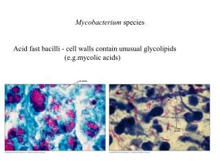
Definition: • Generally, these microorganisms are characterized by their special reaction to stain. • They are difficult to be stained by ordinary stain due to the high content of lipoid substances in the cell wall. • However, once they stained they resist decolorization even with acid, thus they called acid fast bacilli when stained with Zhiel -Neelsen stain.
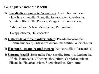
The acid fastness is due to the high lipid content (about 60%) of their cell wall. • They contain N-glycolylmuramic (mycolic) acid instead of N-acetylmuramic acid. • They are Gram +ve but stained poorly. • Non spore forming, non motile and non capsulated. • This group includes both pathogenic and non pathogenic bacteria.
Classification Mycobacterium tuberculosis Mammalian type • Human. • Bovine. • Murin type. M. leprae Atypical Mycobacterium Avian type Reptilian or cold blood type Saprophytic Mycobacterium
Morphology: • Acid Fast beaded bacilli arranged in bundles. • Non spore forming. • Non motile. • Non capsulated. • Culture characteristics: • Require Dorset’s egg media. • Can grow on in presence of Malachite green (Löwenstein-Jensen media) as selective agent.
Strictly aerobic. • Grow very slow, thus require 6-8 weeks before discarding the culture as negative. • Biochemical Reactions: • Niacin test: Positive in human type. Negative in bovine type.
Epidemiology: • Tuberculosis still remains among the most important communicable diseases in the world today with an estimated 50 million active cases of who 3 million will die annually from its effects. • In Egypt, the prevalence rate is about 1.7 % of which two thirds are active cases. The death rate is about 15/100,000.
There are mainly two modes of infection; either by droplet (airborne) infection and this is generally by human type or by consumption of contaminated milk or milk products and this is usually by bovine type. Human type: causes pulmonary tuberculosis mainly by droplet infection from an open case. Bovine type: is attracted by the ingestion of contaminated milk or milk products.
The bacilli will reach to the cervical glands. Also, the organism may reach to the mesenteric lymph nodes causing lymphadenitis. • From the lymph glands the bacilli may reach to many organs causing generalized miliary tuberculosis. • Primary tuberculosis; may occur at any epithelial site (at any part of the body); however, it is most commonly restricted to the lung TB.
Pathogenicity: • Mycobacteria survive after ingestion by macrophages and behave as facultative intracellular organisms. • The infected cells express histocompatibility complex (MHC)-associated bacterial peptides that trigger T cells responses. • Activated CD4+ and TH1 cells release large amounts of interferon- (IFN-), which activates the infected macrophages.
The activated macrophages, in turn, destroy the intracellular Mycobacterium. • The major pathogens are Mycobacterium tuberculosis, the causative agent of tuberculosis, and Mycobacterium leprae, the cause of leprosy. • Atypical mycobacterium, such as Mycobacterium avium-intracellulare complex and Mycobacterium kansasii, can cause tuberculosis disease but less frequent pathogens.
Rapidly growing mycobacteria, such as Mycobacterium chelonei, are saprophytes that occasionally cause human disease in immunocompromised hosts. • In human, the tuberculosis is either: Pulmonary type. Extra-pulmonary type.
Determinants of Pathogenicity: • Cording factor is a glycolipid derivative of mycolic acid that is present on the outer surface of M. tuberculosis. The glycolipid inhibits migration of polymorphonuclear (PMN) leucocytes and elicits granuloma formation. Also, it is immunogenic. • Sulfatides permits the bacteria to survive in the macrophages. • Antibacterial resistance; by mutation.
Clinical picture: • The typical symptoms include; fever, fatigue, night sweats, and weight loss. • Many organs can be involved. Miliary tuberculosis (spread via blood) is characterized by multiple disseminated lesions that resemble millet seeds. • Tuberculous meningitis and tuberculous osteomyelitis are important disseminated forms.
Laboratory Diagnosis: Direct Methods: • The specimen. • Direct Examination (Zhiel-Neelsen stain). Methods Depend on Isolation: • Culture. • Biochemical reactions. • Animal inoculation. Methods other than isolation: • Tuberculin (Mantoux) Test. • Chromatographic Analysis. • Molecular Methods.
Direct Methods: • The specimen: • Sputum: in case of pulmonary tuberculosis. • Pus: in case of skin type. • Urine: in case of urinary tuberculosis. • CSF or blood: in case of meningitis. • Blood: in case of miliary tuberculosis. • Stool: in case of intestinal tuberculosis.
Special Remarks: • For sputum: It's better to take the morning sample and for 3 successive days. The sample requires decontamination process and may be concentration. • Pus: could be treated as sputum. • Urine: is treated as above and in direct examination acid and alcohol must be used to avoid the false +ve result due to presence of the saprophytic mycobacterium in the smegma.
CSF: it's a normal sterile liquid, thus direct examination and culture are done directly from the deposit after centrifugation without the need of decontamination process. • Stool: treated as urine.
Direct Examination: • A film is prepared and suspected specimen and is stained with Ziehl - Neelsen stain. • The appearance of acid-fast bacilli having the morphology of TB (tubercle bacilli) is almost diagnostic of tuberculosis infection. • Alternatively, the bacilli could be detected with auramine stain and visualized by fluorescence microscope.
Methods Depend on Isolation: • Culture: • May be positive even when the direct test is negative and this could be attributed to the presence of very few numbers of bacilli in the sample. • The sample (from non sterile sites) is undergone a process of decontamination and concentration.
Culture is done on Löwenstein-Jensen or Middlebrook medium and incubates aerobically at 37 C and examined weekly up to 8 weeks before considering it negative. • Colonies are dry and warty appearance and buffy in color (human type) or soft and flat colonies (bovine type). • Alternatively, culture could be made in liquid BACTEC medium, in which radioactive metabolites are used and growth can be detected by the production of radioactive CO2 in shorter time.
Biochemical reactions: • Niacin test is +ve in case of human type and -ve in case of bovine type. • Catalase -ve. • Animal inoculation: • The prepared sample for culture may also be injected into two guinea pigs. One of these animals is killed after 4-6 weeks and examined for the typical lesions of tuberculosis and also by film stain and culture. The other animal is left under observation.
Methods other than isolation: • Tuberculin (Mantoux )Test: • The immunological base of this test is the type IV hypersensitivity or delayed type which depends on cell mediated immune reaction. • This test was firstly described by Koch who used the crude extract of TB (tubercle bacilli) known as Koch's Old Tuberculin (K.O.T). • Recently, a purified protein derivative (PPD) is used instead.
Tuberculin test is performed by intradermal injection of the PPD (5 IU) in one forearm. • In case of +ve tuberculin, a red and indurated area (10 mm in diameter) appears after 2-3 days. The induration is very important. • Significance of tuberculin test: • Tuberculin + ve; means a case or previously exposed to TB or vaccinated. Since by adulthood as many as 80% of Egyptians are positive, thus the test is of great diagnostic value (together with the clinical symptoms) in childhood.
Tuberculin -ve; means neither a case nor having immunity. • Tuberculin test is used before BCG vaccination, which should be given only to tuberculin -ve people. If it is given to tuberculin +ve individual, it may provoke a virulent unwanted reaction.
Bacillus Calmette–Guérin is a vaccine against tuberculosis that is prepared from a strain of the attenuated (weakened) live bovine tuberculosis bacillus,Mycobacterium bovis, that has lost its virulence in humans by being specially subculture in an artificial medium for 13 years, and also prepared from Mycobacterium tuberculosis.
Albert Calmette Camille Guérin
Chromatographic analysis • Depend on the analysis of fatty acids by Gas chromatography or HPLC. • Molecular Methods: • These methods are based on the detection of a specific gene(s) of the TB. Of these methods: DNA probe method (Nucleic acid hybridization). • Nucleic acid probes are commercially available. The detection is done within 2-3 hours.
PCR method. • It is a rapid and sensitive method and does not require the presence of the organism but only a specific DNA fragments to be present. • The limitations of these two methods are: • The cost, technical expertise. • The unavailability of the organism to do antibiotic sensitivity testing.
Prevention and Control: • B.C.G. vaccine; it is a living attenuated vaccine derived from M. bovis. Attenuation is obtained by repeated subcultring (about 250 times) on unsuitable medium containing glycerol, potato and bile. • Hygienic measures, etc. • Eradication of infected animals (tuberculin +ve) by slaughtering. • Good nutrition and effective pasteurization of milk.
Treatment: • Because of drug resistance, antibiotic sensitivity test should be performed. • Resistance by mutation.
Atypical or Anonymous MycobacteriumMycobacterium other than tuberculosis (MOTT)
General Features: • Acid fast bacilli differ from the typical TB in being non pathogenic to guinea pig. • They can be treated with antibiotics and they are resistant to anti-tuberculous drugs. • They are widely distributed in the environment. • They form smooth and pigmented colonies. • They are classified according their culture characteristics into:
Atypical or Anonymous Mycobacterium Slow Growing Rapid Growing Photochromogenic Scotochromogenic Non Chromogenic
Slow growing: A. Photochromogenic: • Produce yellow pigment when the growth is being exposed to light. • Example: M. kansasii, M. marinum. B. Scotochromogenic: • Produce orange color chiefly in the dark. • Example: M. scrofulaceum.
C. Non Chromogenic: • Produce no pigments. • Example: M. avium-intracellular complexes.
Rapid growing: • Grow within few days and they can grow in ordinary media. • They can produce several types if infection even mimics TB in symptoms. • Examples: M. fortuitum -chelonei complex. M. smegmatis is a rapidly growing mycobacterium that is not associated with human disease. It is part of the normal flora of smegma, the material that collects under the foreskin of the penis.
Morphology: • They are acid and alcohol fast bacilli when stained with modified Ziehl- Neelsen stain (5% H2 SO4). • They may appear beaded but coarser than TB. • They arrange in masses or groups and mostly intracellular. • Non spore forming. • Non motile.
Culture: • They cannot grow on any artificial media or cell culture. • It can be grown in the mouse footpad or in the armadillo. • Humans are the natural host. • The optimum temperature for growth is 30 °C. It is lower than body temperature; it thus grows preferentially in the skin and superficial nerves.
Epidemiology: • Transmission: Infection is acquired by prolonged contact with patients with lepromatous leprosy, who discharge M. leprae in large numbers in nasal secretions and from skin lesions. • The incubation period is extremely long, lasting from several months to 20 years. • The disease occurs worldwide, with most cases in tropical regions of Asia and Africa.
Pathogenicity: • Leprosy in man occurs in three clinical forms: The nodular or lepromatous type: - In which the organisms produce nodules and form granulation in skin, mucous membrane and internal organs. Tuberculoid type. • In this case the nerve endings are usually affected with paralysis or loss of sensation of the affected area. Mixed type.