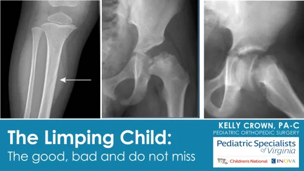
The Limping Child: The good, bad and do not miss
500 likes | 1.73k Views
The Limping Child: The good, bad and do not miss. Kelly Crown , PA-C Pediatric Orthopedic Surgery. Disclosures. I have no relevant financial relationships with the manufacturer(s) of any commercial product(s) and/or provider(s) of commercial services discussed in this CME activity.

The Limping Child: The good, bad and do not miss
E N D
Presentation Transcript
The Limping Child:The good, bad and do not miss Kelly Crown, PA-C Pediatric Orthopedic Surgery
Disclosures • I have no relevant financial relationships with the manufacturer(s) of any commercial product(s) and/or provider(s) of commercial services discussed in this CME activity. • I do not intend to discuss an unapproved/investigative use of a commercial product/device in my presentation
Objectives • Learn to recognize which pediatric limps are concerning and require urgent/emergent care. • Ultimately: When should you be concerned • Review the most common and the most concerning etiologies of pediatric limp • Learn how clinical, radiographic and lab values can help you differentiate between these various etiologies
What is a Limp? • Asymmetric gait • Results in decreased time in the Stance Phase • When it’s painful = Antalgic Gait • Painless: Typically Trendelenburg Gait seen
BIGGEST TAKE AWAY When a child says they have knee pain….. • ALWAYS EXAMINE THE HIPS
Differential Diagnosis All ages: Tumors, Trauma
Septic Arthritis #1 thing you MUST rule this out in all limping kids • Serious bacterial infection within the joint • Delay in treatment cartilage damage, AVN, osteomyelitis • Hematogenus spread • Neonates: Group B Strep, Gram Negative Bacteria • Infants: MSSA, H. Influenza • Children: MSSA, Salmonella • Adolescents: MSSA, Gonorrhea • Can affect any body part and kids of any age but 50% are under the age of 2 • 0-2 y/o Hips 50% • 3+ Knees 50%, Hip 20% SURGICAL EMERGENCY
Septic Arthritis Symptoms PhysicalExam • Refuse to bear weight or limp • Knee or hip pain • Fever • NO injury • No improvement with NSAIDs/Motrin Challenge • Febrile or low grade fever • Toxic/sick looking • Neonates less likely to be toxic • SEVERE pain with passive ROM. • Positive Log Roll (hips) • Asymmetry in motion • Very hesitant to let you examine them • Hip held in flexion and ER • Can have TTP, erythema and when severe, visible effusion
Kocher Criteria Used to identify septic hip arthritis vs. transient synovitis 1. Refusal to bear weight 2. Fever >38.5°C (101.3°F) 3. WBC >12,000 cells/uL 4. ESR >40 mm/h **CRP >20 mg/L or >2mg/dL ** (not part of original criteria, but shown to be most reliable indicator) (J. Bone Joint Surg. Am. 1999;81:1662-70)
Septic Arthritis Tests Treatment • EMERGENT SURGERY for I&D • Sometimes, repease I&D’s are nessesary • continue to follow the CRP • If it’s increasing, take back to surgery • 6 weeks IV antibiotics • CBC, CRP, ESR • *Kocher Criteria • Xray: Pelvis AP/Frog View • Normal doesn’t mean something isn’t wrong. • Late sign is narrowing, edema and ANV/collapse of the femoral head • Ultrasoundif 2+ Kocher Criteria • MRI more sensitive but can delay treatment • Aspiration • >50,000 cells/mm3, >75% segmented neutrophils • Positive gram stain EITHER SURGERY
Transient (Toxic) Synovitis* No. 1 Cause of limp/hip pain in children age 2-8 • Self-limitedinflammatory joint condition • Most commonly affects the hip • Most commonly complain of “Knee/Thigh Pain” • Males 2:1 • Diagnosis of Exclusion • MUST RULE OUT SEPTIC ARTHRITIS • No single laboratory or imaging study will be definitive to confirm or exclude transient synovitis.
Transient (Toxic) Synovitis Symptoms PhysicalExam • Afebrile or low grade fever • Not toxic/sick looking • Passive ROM: Asymmetry +/- guarding and pain • Hips: decreased Abduction and Internal Rotation (+Impingement) • Knee: lack full flexion +/- lack terminal extension • No TTP, No erythema, No visible effusion • Limp or refuse to bear weight • Knee or hip pain • REMOTE history of an ILLNESS • 1 to 5 weeks ago • Viral or bacterial • NO injury • Typically worse in the morning • AFEBRILE or low grade fever (99-101) • Limp improves with NSAIDs
Transient (Toxic) Synovitis Tests Treatment • Self limiting • Symptoms can resolve in a week or as long as 4-5 weeks • The faster the patient is put on Motrin, TID, the faster their pain/symptoms resolve • CBC, CRP, *ESR • Normal • Xray: Pelvis AP/Frog View • Normal vs slight widening • Ultrasound 0.43 0.49
Toddler’s Fractures • Low energy injury • i.e.) Trip and fall over a toy • Classic: going down a slide • Age < 3y/o • Walking toddlers: low likelihood of child abuse • If not walking, suspect abuse • STABLE fractures: commonly spiral/oblique fracture of the tibia • Other common locations • 1st Metatarsal • Tibia +/- Fibula • Femur (higher suspicion for abuse)
Toddler’s Fractures Symptoms PhysicalExam • Afebrile, Nontoxic • BONY TTP and/or pain with rotation • Passive ROM: Asymmetry +/- guarding and pain • Pain with hip ROM: femur • Pain with knee ROM: distal femur or proximal tibia • Pain with ankle ROM: distal tibia/fibula • Limp or refuse to bear weight • Crawling: tibia, fibula fx • Not walking: tibia or femur fx • Walk on lateral aspect of foot or on their heel: Metatarsal fx • Injury • AFEBRILE
Toddler’s Fractures Tests Treatment • Self limiting • Cast vs observation • Heal in 2-4 weeks • Limp up to 6 weeks • Typically no long term sequela • Proximal Tibia (Cozen Fracture) slight risk of developing Cozen Deformity (Valgus deformity of the knee 1-2 years after) • Xray: nondisplaced subtle fracture • Sometimes a clinical diagnosis • Might not be visible for a week • Late finding is new periosteal reaction
Leg-Calve-Perthesaka Perthes • Avascular Necrosis of the Femoral Head • Unknown etiology • Can cause permanent deformity of the femoral head • Earlier treatment, minimizes this risk of permanent deformity • Boys 5:1 more likely • Age typically 4-7 • Age <6 better prognosis • Age > 8 worse prognosis
Leg-Calve-Perthes Symptoms PhysicalExam • PAINLESS LIMP (first symptom) • +/- Hip, thigh or Knee • NO injury • Afebrile Symptoms Wax and Wane • Worse during or after activities • Improve with rest and NSAIDs • Antalgic gait • Trendelenburg gait late finding once femoral head collapses • Decreased hip ROM • Loss of Abduction and Internal Rotation • Hip flexion contracture • Limp Length Discrepancy (late finding) • Hip contracture can exacerbate this
Leg-Calve-Perthes Tests Treatment **type of treatment recommended is based on patient’s age, stage of disease, and radiographic class** MAIN GOALS • Symptomatic relief: NSAIDs • Protected weight bearing through the acute phase with crutches +/- abduction brace Can take 18mo to 2 years to fully subside • Xray • Normal first 3-6mo • Early signs is medial joint space narrowing, sclerosis and flattening of the femoral head • MRI is the gold standard for diagnosis • Follow with serial xrays
SCFE(Slipped Capital Femoral Epiphysis) • Femoral head Slips off the Femoral neck • “Ice cream falling off the cone” • Most common adolescent hip disorder • During rapid growth • Males: 13.4 y/o (12-16) • Females 12.2 y/o (10-14) • Younger: Hypothyroid • **Obesity** • Male (2 to 1.5 ratio) • Unilateral (40% BL) SURGICAL EMERGENCY Jorge Muniz, PA-C, Medcomic.com
SCFE Symptoms PhysicalExam • Gait • Antalgic gait = Stable SCFE • NWB = Unstable SCFE • Decreased hip ROM • Loss of Internal Rotation • Obligatory ER with hip flexion • Guarding • Limp Length Discrepancy (unstable) • Short and Externally rotated • PAINFUL LIMP • Acute or chronic • Hip pain (L>R) • Commonly get referred thigh or knee pain • +/- minor injury • Afebrile
SCFE Tests Treatment • Percutaneous Screw Fixation • URGENT • Contralateral Pinning? • Correct the deformity after it heals • Minimal displacement: hip arthroscopy with chondroplasty (FAI surgery) • Displaced: Varus Derotational Osteotomy (VDRO) • Xray Pelvis • AP & Frog-leg Lateral • Frog-leg best at subtle SCFE • MRI if nlXray: can dx pre-slip • Physeal edema or widening
Case 1: Left leg pain x 13 days. Hasn’t walked in 12 days 7 y/o Female No PMH 2 weeks ago fell from a seated position off a rocking chair. Walked and had no pain after the fall. Next morning woke up with left knee pain and wouldn’t walk 2 days later, had a tactile fever. Taken to local ER, Temp 100.4. Hip Xray Slight Narrowing. Positive for StrepAand put on Amoxicillin Day 7 of her ABX develops a rash, back to urgent care. Still not walking! Temp 99. Thought to be a drug rash. Stopped the Amoxicillin and put on Ceftin. • CBC: elevated WBC (13.6), Anemic, Hgb 10.5, Hct 31.1, Platelets 724 PCP instructed the family OK not to take ABX since 7 days was sufficient
Case 1 • Sees PCP the AM before coming to the ortho clinic, order labs • CBC: nl WBC (10), worsening anemia Hgb 9.1, Hct 28, Platelets 671 • Sed Rate 95 DAY 13: Sees me • HASN’T WALKED IN 12 DAYS. • Parents still say that tactilely she is still febrile (don’t have a thermometer) • No improvement in pain with Motrin or Tylenol. • Any small movement, she cries in pain.
Case 1 • Vitals • Temp 98.2 F (took motrin 2 hrs prior) • Unable to get height and weight because she refuses to stand • PE • Sitting in a baby stroller • Looks sick • SEVERE pain with even minor left hip ROM • Hip flexion contracture with obligatory ER • Full knee ROM without pain • TTP at Psoas
Case 1 • Admitted to Fairfax ED for STAT MRI with aspiration • Labs • WBC 14.12 4.80 - 13.00 x10 3/uL • Hgb9.5 11.5 - 14.5 g/dL • Hematocrit 29.0 33.0 - 43.0 % • Platelets681140 - 400 x10 3/uL • CRP 6.3 0.0 - 0.8 mg/dL • ESR99 0 - 20 mm/Hr
Kocher Criteria Used to identify septic hip arthritis vs. transient synovitis 1. Refusal to bear weight 2. Fever >38.5°C (101.3°F) 3. WBC >12,000 cells/uL 4. ESR >40 mm/h **CRP >20 mg/L or >2mg/dL ** (not part of original criteria, but shown to be most reliable indicator) (J. Bone Joint Surg. Am. 1999;81:1662-70)
Case 1 • MRI • Septic arthritis with Psoas Abscess • URGENT I&D • Cartilage of her hip was gone, underlying osteomyelitis • 6 weeks IV ABX
3 months post-op Still has pain and limited hip ROM
Case 2: Left hip pain x 2 weeks • PE: • Vitals • 5’3” 130lbs • Afebrile • AAO x 3, NAD • Slightly antalgic gait but FWB • TTP at the groin • Pain and Limited IR 11 y.o. female No PMH • Left hip pain x 2 weeks after a fall • jumping over hurdles at camp and landed on her LLE "awkwardly." • Pain= sharp, non-radiating, worse with movement • Able to walk
Case 2 • URGENT Percutaneous Screw Fixation • 7 weeks post-op • Pain free • FWB • Only lacked 10 degrees IR • Full abduction • CLEARED FOR ALL ACTIVITIES
Case 3: 1 year of left knee pain 16 y/o M with no PMH here for 2nd opinion • Left knee pain x 1 year after a twisting injury during soccer • Didn’t get treatment for 2 months • Was seen by an outside ortho group for concern for meniscus tear • MRI showed small lateral meniscus tear • Tx included rest, NSAIDs, corticosteroid injection and months of physical therapy • Physical Therapist noted that he had minimal hip ROM • Has had no hip pain x 2 months
Case 3 Vitals • 5’7” 187lbs • Afebrile PE • IR of the Right was 40, Left was 0 and with mild pain • Obligate ER of the left • Full knee ROM, negative McMurry • Gait: Significant Trendelenburg and External Foot progression of the left
Case 3 TREATMENT Option 1: • Percutaneous Screw Fixation • POSITVE: would stop further Slippage, minor surgery, WBAT after • NEGATIVE: wouldn’t correct the deformity, likely need a second surgery Option 2: • VDRO (Valgus Derotational Osteotomy) • POSITIVE: Would correct the deformity and regain his motion • NEGATIVE: Major surgery, would be NWB for 4-6 week and not able to run for 12 weeks
They choose option 210 weeks post-op -now FWB-knee pain -recently started PT to help with his hip ROM and strength now that the osteotomy is healed
BIGGEST TAKE AWAY When a child says they have knee pain….. • ALWAYS EXAMINE THE HIPS
Thank you! Questions?


