Techniques in Protein Biochemistry
370 likes | 575 Views
Techniques in Protein Biochemistry. Chapter 5. Problem: isolation & analysis of protein or amino acid found in cell Assumption: can somehow analyze for wanted protein Common – Colorimetric indicator (chemical rxn color form’n; can be monitored spectrophotometrically)

Techniques in Protein Biochemistry
E N D
Presentation Transcript
Techniques in Protein Biochemistry Chapter 5
Problem: isolation & analysis of protein or amino acid found in cell • Assumption: can somehow analyze for wanted protein • Common – Colorimetric indicator (chemical rxn color form’n; can be monitored spectrophotometrically) • Functional indicator (biological endpoint) • This example – colorimetric (breakdown of fats purple color) • Activity assay • Use at each step of separation
Isolation of Wanted Protein from Brain Cells • Brain cells contain wanted protein • Open cells • Homogenization, sonication, grinding • Maintain cold, pH, osmolality • Centrifugation often used Known speeds/ conditions for different organelles
Test each fraction for activity • Save most active fractions • Separation of wanted protein from other types of molecules • Dialysis against physiological buffer
Separation from other proteins • ChromatographyAll use solid or aqueous support to which wanted protein has some affinityAll use aqueous or gaseous mobile phase; wanted protein has different affinityThis also moves molecules through/ past support
If wanted protein has greater affinity for support than for mobile phase, protein “adheres” to support phase • If wanted protein has greater affinity for mobile phase than for support, protein will move with mobile phase through/away from support
Gel Filtration Chromatography (= Size Exclusion) • Separation by MW • Solid support = porous beads (ex: sephadex, sepharose) • Held in column • Beads have microscopic pores/pits/spaces • Mobile phase = buffer of physio pH, ionic strength
Sample = sol’n of wanted protein + other (unwanted) proteins; most have different MWs • Apply sample to column • Begin slow mobile phase flow • Smaller proteins enter spaces in beads • Larger proteins flow w/ buffer around beads (so emerge 1st from column) • Collect fractions; test each fraction by activity assay
Ion Exchange Chromatography • Separation by overall charge of proteins • Solid support = resin (charged microscopic beads) suspended in buffer • Mobile phase = buffer of particular pH, ionic strength • Sample = sol’n of wanted protein + other (unwanted) proteins Most have different overall + or – charges of various strengths
Apply sample to column, begin slow mobile phase flow • Proteins of charge opposite that of resin + of similar strength of charge of resin: Good affinity for resin; bind electrostatically • Proteins of the same charge or different strength of charge of resin: No good affinity for resin; flow through column quickly, so eluted first • Result: protein similar to resin is held in column
To elute protein held to resin in column: use buffer of higher ionic strength or stronger pH • Changes ionic environment • Ions in new (elution) buffer “exchange” for protein (are more attractive to resin, so take the place of protein on resin) • Collect fractions from mobile phase + elution buffer • Test all fractions by activity assay
Affinity Chromatography • Based on specificity of prot of interest for some molecules to which it alone will bind • Ex: Ab binds only specific Ag • BUT binding must be reversible • Solid support = specific binding molecule (=ligand) covalently bound to beads, etc. • Mobile phase = buffer of proper pH, ionic strength to maintain activity of prot of interest
Pack column; apply sample • Begin slow mobile phase flow • Prot of interest ONLY will bind to ligand • Types of binding (must be reversible): ionic, H-bonds, hydrophobic interactions • To elute, may use solution of ligand (competes w/ solid phase ligand) OR buffers of diff strength, pH that disrupt protein/ligand interactions
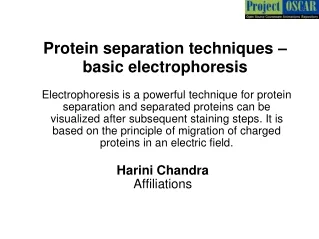
Electrophoresis • Separation AND identification • Based on overall charge of protein movement under influence of electric field • Zone • Semisolid or gelatinous medium (plate or slab) • Spot protein mixture (w/ wanted + unwanted proteins in solution) onto gel • Apply electric field
Molecules migrate toward anode (+ charged) • Distance traveled dependent on charge, size of protein Most impt = size of protein • Gel support acts as molecular sieve; smaller molecules go faster toward anode, so migrate further • Also, those more strongly charged move closer to anode • Use chemical to stain aa’s bands representing proteins of decreasing MW
Run stds simultaneously • Mixture of proteins of known MW; spot on one or several lanes • Electrophorese under same conditions as unknown protein mixture • Stain “ladder” of bands (lowest to highest MW proteins traveling some distance under these conditions)
Determine distance traveled for each band from origin • Plot x = distance migrated for each std of known MW; y = log MW of stds • Yields std curve; find distance traveled by unknown prot(s) on curve; deter MW • Can also cut gel, dissolve to free proteins
Moving Boundary = IsoElectric Focusing (IEF) • Separation due to charge; based on isoelectric pt of each prot • Use gel of ampholytes (gel has regions of different pHs) • Spot sample in middle of gel • Apply electric field
Each prot in mixture will migrate toward + or – electrode, according to charge • Each prot will stop moving when it reaches pH region of gel = its isoelectric pH • Stain aa’s of prot’s w/ chemical
Run stds simultaneously • Mixture of proteins of known pI’s; spot in one or several lanes • Electrophorese under same conditions as unknown protein mixture • Stain “ladder” of bands (lowest to highest MW proteins traveling some distance under these conditions) • Determine distance traveled for each band from origin • Plot x = distance migrated for each std of known pI y = pH • Yields std curve; can find distance traveled by unknown prot(s) on curve pI
Characterize wanted protein by aa sequence • Once wanted protein has been isolated from all other cell molecules • Old method: Break all peptide bonds solution of aa’s • Analyze aa’s by chromatography • Thin Layer Chromatography (TLC) – on coated plate or paper support; various mobile phases separate aa’s from each other • High Pressure Liquid Chromatography (HPLC) – force sample through small column packed with various types of support; various mobile phases are forced through column by high pressure pumps to separate aa’s from each other
Now have identified all aa’s in protein • With original protein, use chem. rxn to label amino terminal aa • Use various enz’s to cleave prot at partic aa’s along peptide chain peptide fragments • Analyze fragments for overlap; use knowledge of all aa’s in protein sequence
New method: Automated • Chem rxn to label amino terminal aa • Cleave amino terminal aa • Analyze for identity of last aa • Rest of prot now has diff amino terminal aa (second to last in original prot) • Chem rxn to label second to last aa of amino terminal • Cleave this terminal aa • Analyze for identity of second-to-last aa • Etc. etc. etc.
Common now: identify gene for protein of interest • Isolate mRNAs (found w/in cell rich in wanted protein) w/ gene nucleotide sequence • Use mRNAs to identify gene • mRNA will have complimentary sequence to gene in DNA, so will pair in that region of DNA only • Analyze gene for nucleotide sequence • Use genetic code to determine aa sequence of wanted protein from gene which codes for it