Download
1 / 70
770 likes | 1.16k Views
Evaluation of the Neck Mass. Adapted from Michael Underbrink, MD Byron J. Bailey, MD. Introduction. Common clinical finding All age groups Very complex differential diagnosis Systematic approach essential. Anatomical Considerations. Prominent landmarks Triangles of the neck

E N D
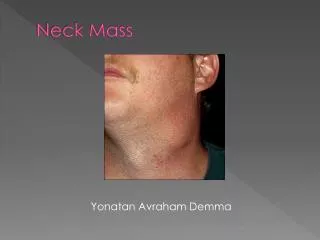
Evaluation of the Neck Mass Adapted from Michael Underbrink, MD Byron J. Bailey, MD
Introduction • Common clinical finding • All age groups • Very complex differential diagnosis • Systematic approach essential
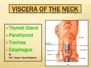
Anatomical Considerations • Prominent landmarks • Triangles of the neck • Carotid bulb • Lymphatic levels
Anatomical Considerations • Prominent landmarks • Triangles of the neck • Carotid bulb • Lymphatic levels
Anatomical Considerations • Prominent landmarks • Triangles of the neck • Carotid bulb • Lymphatic levels
Anatomical Considerations • Prominent landmarks • Triangles of the neck • Carotid bulb • Lymphatic levels
General Considerations • Patient age • Pediatric (0 – 15 years): 90% benign • Young adult (16 – 40 years): similar to pediatric • Late adult (>40 years): “rule of 80s” • Location • Congenital masses: consistent in location • Metastatic masses: key to primary lesion
Risk Factors for Head and Neck Cancer • Chronic sun exposure • Tobacco and alcohol use • Poor dentition • Industrial or environmental exposures • Family history
Symptoms of Head and Neck Cancer • Nonhealing ulcer within the oral cavity or oropharynx • Persistent sore throat • Dysphagia • Change in voice • Recent weight loss
Diagnostic Steps • History • Developmental time course (age, size, acute vs chronic) • Associated symptoms (fever, sore throat, cough, dysphagia, otalgia, voice) • Personal habits (tobacco, alcohol) • Recent travel, trauma, insect bites, pets/farm animals • Previous irradiation or surgery
Physical Exam • Complete head and neck exam • Skin, otologic examination, oropharynx, tongue • Ulcerations, submucosal swelling, or asymmetry, particularly in the tonsillar fossa • Examination of the larynx and pharynx is accomplished by indirect or flexible laryngoscopy. • Palpation during swallowing or during a Valsalva's maneuver may identify pathology within the larynx and thyroid gland. • Rotation of the head in both flexion and extension aids examination of the posterior triangle of the neck.
Empirical Antibiotics • Inflammatory mass suspected • Two week trial of broad spectrum antibiotics • Follow-up 1-2 weeks for further investigation
Diagnostic Tests • Fine needle aspiration biopsy (FNAB) • Computed tomography (CT) • Magnetic resonance imaging (MRI) • Ultrasonography • Radionucleotide scanning
Fine Needle Aspiration Biopsy • Standard of diagnosis • Indications • Any neck mass that is not an obvious abscess • Persistence after a 2 week course of antibiotics • Progressive growth, supraclavicular, > 3 cm • Any symptoms associated with lymphoma • Small gauge needle • Reduces bleeding • Seeding of tumor – not a concern • No contraindications (vascular ?)
Fine Needle Aspiration Biopsy • Proper collection required • Minimum of 4 separate passes • Skilled cytopathologist essential • On-site review best
Computed Tomography • Distinguish cystic from solid • Extent of lesion • Vascularity (with contrast) • Detection of unknown primary (metastatic) • Pathologic node (lucent, >1.5cm, loss of shape) • Avoid contrast in thyroid lesions
Magnetic Resonance Imaging • Similar information as CT • Better for upper neck and skull base • Vascular delineation with infusion
Ultrasonography • Less important now with FNAB • Solid versus cystic masses • Congenital cysts from solid nodes/tumors • Noninvasive (pediatric)
Ultrasonography YROID ASS
Radionucleotide Scanning • Salivary and thyroid masses • Location – glandular versus extra-glandular • Functional information • FNAB now preferred for for thyroid nodules • Solitary nodules • Multinodular goiter with new increasing nodule • Hashimoto’s with new nodule
Nodal Mass Workup in the Adult • Any solid asymmetric mass MUST be considered a metastatic neoplastic lesion until proven otherwise • Asymptomatic cervical mass – 12% of cancer • ~ 80% of these are SCCa
Nodal Mass Workup in the Adult • Ipsilateral otalgia with normal otoscopy – direct attention to tonsil, tongue base, supraglottis and hypopharynx • Unilateral serous otitis – direct examination of nasopharynx
Nodal Mass Workup in the Adult • Panendoscopy • FNAB positive with no primary on repeat exam • FNAB equivocal/negative in high risk patient • Directed Biopsy • All suspicious mucosal lesions • Areas of concern on CT/MRI • None observed – nasopharynx, tonsil (ipsilateral tonsillectomy for jugulodigastric nodes), base of tongue and piriforms • Synchronous primaries (10 to 20%)
Nodal Mass Workup in the Adult • Open excisional biopsy • Only if complete workup negative • Occurs in ~5% of patients • Be prepared for a complete neck dissection • Frozen section results (complete node excision) • Inflammatory or granulomatous – culture • Lymphoma or adenocarcinoma – close wound
Thyroid mass Lymphoma Salivary tumors Lipoma Carotid body and glomus tumors Neurogenic tumors Primary Tumors
Thyroid Masses • Leading cause of anterior neck masses • Children • Most common neoplastic condition • Male predominance • Higher incidence of malignancy • Adults • Female predominance • Mostly benign
Thyroid Masses • Lymph node metastasis • Initial symptom in 15% of papillary carcinomas • 40% with malignant nodules • Histologically (microscopic) in >90% • FNAB has replaced USG and radionucleotide scanning • Decreases # of patients with surgery • Increased # of malignant tumors found at surgery • Doubled the # of cases followed up • Unsatisfactory aspirate – repeat in 1 month
Lymphoma • More common in children and young adults • Up to 80% of children with Hodgkin’s have a neck mass • Signs and symptoms • Lateral neck mass only (discrete, rubbery, nontender) • Fever • Hepatosplenomegaly • Diffuse adenopathy
Lymphoma • FNAB – first line diagnostic test • If suggestive of lymphoma – open biopsy • Full workup – CT scans of chest, abdomen, head and neck; bone marrow biopsy
Salivary Gland Tumors • Enlarging mass anterior/inferior to ear or at the mandible angle is suspect • Benign • Asymptomatic except for mass • Malignant • Rapid growth, skin fixation, cranial nerve palsies
Salivary Gland Tumors • Diagnostic tests • Open excisional biopsy (submandibulectomy or parotidectomy) preferred • FNAB • Shown to reduce surgery by 1/3 in some studies • Delineates intra-glandular lymph node, localized sialadenitis or benign lymphoepithelial cysts • May facilitate surgical planning and patient counseling • Accuracy >90% (sensitivity: ~90%; specificity: ~80%) • CT/MRI – deep lobe tumors, intra vs. extra-parotid • Be prepared for total parotidectomy with possible facial nerve sacrifice
Carotid Body Tumor • Rare in children • Pulsatile, compressible mass • Mobile medial/lateral not superior/inferior • Clinical diagnosis, confirmed by angiogram or CT • Treatment • Irradiation or close observation in the elderly • Surgical resection for small tumors in young patients • Hypotensive anesthesia • Preoperative measurement of catecholamines
Lipoma • Soft, ill-defined mass • Usually >35 years of age • Asymptomatic • Clinical diagnosis – confirmed by excision
Neurogenic Tumors • Arise from neural crest derivatives • Include schwannoma, neurofibroma, and malignant peripheral nerve sheath tumor • Increased incidence in NF syndromes • Schwannoma most common in head & neck
Schwannoma • Sporadic cases mostly • 25 to 45% in neck when extracranial • Most commonly between 20 and 50 years • Usually mid-neck in poststyloid compartment • Signs and symptoms • Medial tonsillar displacement • Hoarseness (vagus nerve) • Horner’s syndrome (sympathetic chain)
Congenital and Developmental Mass • Epidermal and sebaceous cysts • Branchial cleft cysts • Thyroglossal duct cyst • Vascular tumors
Epidermal and Sebaceous Cysts • Most common congenital/developmental mass • Older age groups • Clinical diagnosis • Elevation and movement of overlying skin • Skin dimple or pore • Excisional biopsy confirms