Download
1 / 48
550 likes | 848 Views
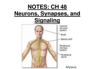
Neurons, Synapses, and Signaling. CHAPTER 48 and 50. Figure 48.1 Overview of a vertebrate nervous system. NERVOUS SYSTEM. Central nervous system (CNS) – brain and spinal cord Peripheral nervous system (PNS) – nerves that communicate motor and sensory signals between CNS and rest of body.

E N D
Neurons, Synapses, and Signaling CHAPTER 48 and 50
NERVOUS SYSTEM • Central nervous system (CNS) – brain and spinal cord • Peripheral nervous system (PNS) – nerves that communicate motor and sensory signals between CNS and rest of body
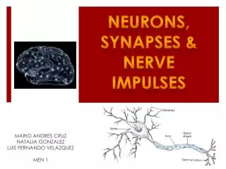
NEURON • Functional unit of nervous system • Relatively large cell body • Processes: • Dendrites – convey signals from tips to cell body; often branched • Axons – conduct signals away from body and toward tip; often single • Myelin sheath – protective, insulating layer that covers many axons in vertebrates • Made by Schwann cells in the PNS • Made by oligodendrocytes in the CNS
Axon ends at synaptic terminals • Synapse – site of contact between synaptic terminal and target cell (neuron or effector cell – for example a muscle cell) • Neurotransmitter – chemical messengers between neurons and other cells
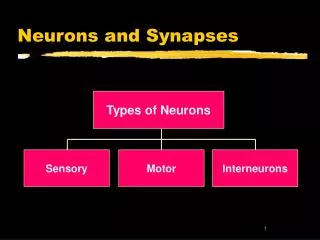
ORGANIZATION OF NEURONS • Sensory neurons – communicate sensory information from eyes and other senses and internal conditions • Senses, blood pressure, muscle tension, CO2 levels) • Interneurons – integrate sensory input and motor output; communicate only between neurons; make up vast majority of brain neurons • Motor neurons – convey impulses from CNS to effector cells (muscles and glands)
MEMBRANE POTENTIAL • Voltage measured across the membrane (like a battery) • Inside of cell more negative • Typically –50 to –80 mV (resting potential) • Sodium-potassium pump keeps ionic gradient (3Na+ out, 2K+ in)
Figure 8.15 The sodium-potassium pump: a specific case of active transport
Charges Across Membranes • Neurons have ability to generate changes in their membrane potential • Resting potential – membrane potential of cell at rest (-60mV to -80mV) • Gated ion channels control membrane potential – open to different stimuli • Hyperpolarization – increase in electrical gradient • Open K+ channel (K+ moves out) • Cell becomes more negative • No action potential because it makes it harder to depolarize
Depolarization – decrease in electrical gradient • Open Na+ channel (Na+ moves in) • Cell becomes more positive • Action potential generated if threshold is reached (-50mV to -55mV) • Massive change in voltage • Threshold causes all-or-none event • Action potential - massive change in membrane voltage that can spread along the membrane
Figure 48.8 Graded potentials and the action potential in a neuron
Figure 48.9 The role of voltage-gated ion channels in the action potential
ROLE OF GATED CHANNELS • Depolarizing – Na+ gates open rapidly so Na+ moves into cell • Repolarizing – K+ gates finally open and K+ moves out; Na+ gates close • Undershoot (Refractory Period) - K+ still open (they are slower to close) and Na+ still closed so cell becomes even more negative than resting and cannot be depolarized • Stronger stimuli result in greater frequency of action potentials and NOT from stronger action potentials • Propagation • Action potentials move in one direction due to refractory period
Propagation of the action potential Na+ moves into cell starting action potential. Depolarization spreads and K+ repolarizes initial area. Prevents action potential on that side.
Figure 48.11 Saltatory conduction • Voltage leaps from node to node
SYNAPSES • Presynaptic cell – transmitting cell • Postsynaptic cell – receiving cell • Two types of synapses • Electrical • Need gap junctions (channels between neurons) • No delays • Chemical • Narrow gap, synaptic cleft, between cells • More common than electrical in vertebrates and most invertebrates • Require neurotransmitters (chemical intercellular messengers)
Depolarization of presynaptic membrane causes influx of Ca2+ • Increased Ca2+ in cell causes synaptic vesicles to fuse to cell membrane and release neurotransmitters via exocytosis • Neurotransmitters diffuse to postsynaptic cell • Postsynaptic membrane has gated channels that open when neurotransmitters bond to specific receptors
A single neuron may receive many inputs simultaneously • Neurotransmitters cause 2 different responses depending on the gates that are opened • Inhibitory • (hyperpolarization) • Excitatory • (depolarization) • Neurotransmitters are quickly degraded • Excitatory postsynaptic potential (EPSP) – Na+ in and K+ out = depolarization • Inhibitory postsynaptic potential (IPSP) - K+ out or CL- in = hyperpolarization
NEUROTRANSMITTERS • Acetylcholine • one of the most common • can excite skeletal muscle and inhibit cardiac muscle • Epinephrine and norepinephrine • also function as hormones
Dopamine • Usually excitatory • Excess dopamine can cause schizophrenia • Lack of dopamine can cause Parkinson’s • Sertonin • Usually inhibitory • Endorphins • Natural painkillers (morphine and opium mimic endorphins shape) • Nitric Oxide (NO) • Released during sexual arousal (increasing blood flow) • Nitroglycerin used to treat chest pain
SKELETAL MUSCLE • Attached to bones and responsible for their movement • Consist of bundles of long fibers • Each fiber is a single cell with many nuclei
Each fiber made up of smaller myofibrils • Myofibrils made of 2 kinds of myofilaments • Thin myofilaments • 2 strand of actin with a regulatory protein (tropomyosin) • Thick myofilaments • Staggered arrays of myosin
Striated muscle due to repeating light and dark bands • Sarcomere – basic unit of muscle • Contraction of sarcomeres results in muscle contraction. • Actin and myosin slide pass each other to shorten the sarcomere.
Figure 49.32 The sliding-filament model of muscle contraction
Figure 49.33 Myosin-actin interactions generate the force for muscle contraction
Sliding-filament model • Myosin head phosphorylated by ATP making the head energized • Energized head attaches to actin making cross-bridge • ADP and Pi released from head so it goes back to relaxed state, sliding the thin filament toward center of sarcomere • A new ATP binds to head releasing it from actin • Creatine phosphate – stores phosphate in vertebrate muscles
How is skeletal muscle contraction regulated? • An action potential begins in the brain and travels via nerve to muscle. • The action potential causes neuron to release acetylcholine (neurotransmitter). This results in an excitatory response in muscle.
Acetylcholine triggers action potential in T-tubuleswithin muscle • T-tubulesare infoldings of muscle cell’s cell membrane
T-tubules touch sarcoplasmic reticulum and change is permeability to Ca2+ which means it releases Ca2+ • Sarcoplasmic reticulum– specialized ER that stores Ca2+ • Ca2+ binds to troponin which frees binding site for myosin head
Figure 49.35 The roles of the muscle fiber’s sarcoplasmic reticulum and T tubules in contraction
What’s troponin and tropomyosin? • Tropomyosin blocks myosin heads binding sites • Troponin controls position of tropomyosin • When Ca2+ binds to troponin, the shape of tropomyosin-troponin complex changes and frees binding site
Figure 49.34 Hypothetical mechanism for the control of muscle contraction
Summation and frequency of action potentials determine muscle tension • One muscle cell only innervated by one motor neuron, but one motor neuron may innervated many muscle cells • More cells activated = more tension
Big Picture – Making a muscle contract • Action potential generated in brain and travels down nerve • Action potential causes acetylcholine to diffuse across synapse to muscle • Acetylcholine causes excitatory responses (action potential) that moves down T-tubules • Change in membrane potential causes SR to release calcium • Calcium binds to troponin, which then moves tropomyosin • ATP used to bind myosin head to actin • Sarcomere contracts and then ATP used to break bridge