Download
1 / 64
660 likes | 1.73k Views
The Sensory Organs. 山东大学医学院 解剖教研室 李振华. The Sensory Organs. Sensory organs include the receptors and accessory organs . The receptors may be divided into three kinds: The exteroceptors 外感受器 : receive stimuli such as touch, temperature, pain, light and sound from the external environment

E N D
The Sensory Organs 山东大学医学院 解剖教研室 李振华
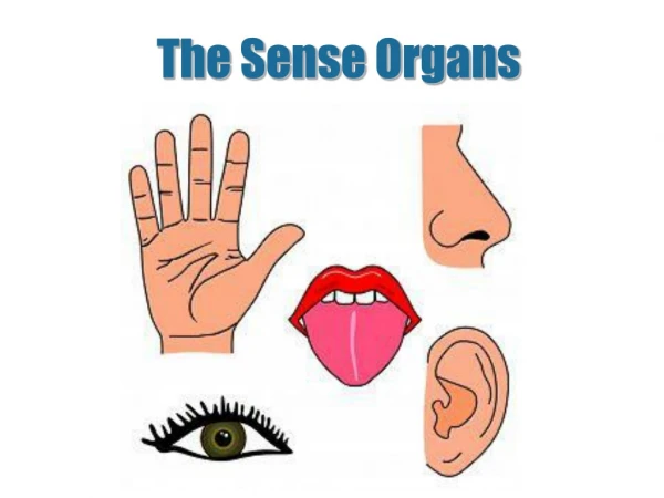
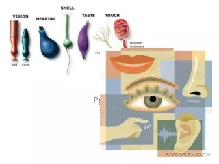
The Sensory Organs Sensory organs include the receptors and accessory organs. The receptors may be divided into three kinds: • The exteroceptors外感受器: receive stimuli such as touch, temperature, pain, light and sound from the external environment • The interoceptors 内感受器: pick up information about internal environment • The proprioceptors 本体感受器: receive stimuli from muscles, tendons, joints and ligaments
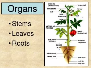
The Visual Organ 视器 Composition: eyeball and accessory organs of eye Shape of eyeball • Has anterior and posterior poles • Equator 赤道: an imaginary line encircling the eyeball, midway between anterior and posterior poles • Axis of eyeball眼轴: a line joining the two poles • Optic axis 视轴: a line joining the center of the pupil to the fovea centralis
Walls of eyeball Cornea 角膜 Fibrous tunic of eyeball Sclera巩膜 Iris 虹膜 Vascular tunic of eyeball Cilliary body 睫状体 Choroid 脉络膜 Pars iridica retinae Pars caeca retinae Retina 视网膜 Pars ciliaris retinae Pars optica retinae
Fibrous tunic of eyeball (outer) • Cornea 角膜: anterior 1/6, a nonvascular, transparent portion, richly supplied by nerves; because it is curved, the cornea helps focus light • Sclera巩膜(white of eye, opaque portion): • posterior 5/6, consisting of fibrous connective tissue that forms a tough protective covering for eyeball, • Contains sinus venosus sclerae巩膜静脉窦which lies beneath the junction of cornea and sclera
Vascular tunic of eyeball (middle) • Iris 虹膜 • Thin contractile membrane anterior to ciliary body, with a central opening, the pupil 瞳孔 • Contains sphincter pupillae瞳孔括约肌(circular fibers) and dilator pupillae瞳孔开大肌 (radial fibers) • Cornea and iris meet to form the iridocorneal angle 虹膜角膜角
Cilliary body 睫状体 • Body a ring-shaped thickening anterior to equator, containing smooth muscle fibers called ciliary muscle 睫状肌 • Ciliary processes 睫状突: a series of some 60~80 projections producing aqueous humor • Ciliary ring 睫状环
Sinus venosus sclerae Ciliary Muscle Iridocorneal angle Dilator Pupillae Sphincter Pupillae Lens ciliary zonule Ciliary Processes Ciliary ring
Choroid 脉络膜 • Thin, highly vascular in posterior 2/3 of eye • Contains brown pigmented cells and dense capillary plexus
Retina 视网膜 • Pars caeca retinae视网膜盲部 • Pars iridica retinae 视网膜虹膜部 • Pars ciliaris retinae 视网膜睫状体部 • Pars optica retinae视网膜视部 • Lines the choroidsComposed of two layers • An outer pigment cell layer • Inner neural layer (four layers)
Ganglion cells Bipolar neurons Rod cells Cone cells Pigment cell layer • The fourth layer consists photoreceptor cells • Cone cells视锥细胞are color receptors that function best during the day • Rod cells视杆细胞are dark-light receptors that function best at night and in dim light • The third layer consists of bipolar neurons 双极细胞 • The second layer is formed ganglion cells 节细胞, whose axons form optic nerve • The first layer consists of nerve axons that collect at the optic disk and pass through the sclera to form the optic nerve
Optic disc 视神经盘(blind spot), located medial to posterior pole of eye, and consists of optic nerve fibers and central artery of retina • Macula lutea 黄斑 • Lies lateral about 3.5 mm to optic disc, a shallow depression, it is completely free of blood vessels and is yellowish in color • Fovea centralis 中央凹, aera of greatest visual acuity (concentration of cones), at its center • The pigmentted layer absorbs light that passes completely through the anterior layer, preventing backscatter (blurring of vision)
Contents of eyeball Aqueous humor 房水 Lens 晶状体 Vitreous body玻璃体
Aqueous humor 房水 Chamber of eye 眼房-lies between cornea and lens, and divided by iris into anterior and posterior chambers Aqueous humor 房水 • A clear watery fluid that fills chamber of eye • Continuously secreted by ciliary body into posterior chamber • Passes through pupil into anterior chamber • Then it filters though iridocorneal angle into sinus venosus sclerae, this sinus drains via anterior ciliary veins into ophthalmic veins
Pupil Iridocorneal angle Production and circulation of aqueous humor Ciliary body Posterior chamber Anterior chamber Sinus venosus sclera Anterior ciliary vein Ophthalmic vein Functions • Helps focus light • Helps maintain constant pressure in eyeball • Helps nourish the lens and cornea
Lens晶状体 • Transparent biconvex structure, covered by an elastic transparent capsule • Located between iris and vitreous body, and suspended behind pupil by ciliary zonule 睫状小带 • Shape changed by the ciliary muscle: for near vision, the ciliary muscle contracts and the lens rounds up, while for distant vision the lens flattens out, so that the eye may be focused on distant objects
Vitreous body 玻璃体 • Consists of colorless, transparent jelly-like substance in which there is a meshwork of fine fibrils, occupies the vitreous chamber, the space between lens and retina • Helps maintain the shape of eyeball and supports the retina
Refractive media of eye折光装置 Bend entering light waves and focus them on the retina • Cornea • Aqueous humor • Lens • Vitreous body
Accessory organs of eye 眼副器 Eyelids 眼睑 Conjunctiva 结膜 Lacrimal apparatus泪器 Ocular muscles眼球外肌 Connective tissue in the orbit
Eyelids 眼睑 (from without inwards ) • Skin, extremely thin • Subcutaneous areola tissue, loose and delicate • Muscular layer: orbicularis oculi • Tarsus睑板, formed by dense connective tissue in which the tarsal glands睑板腺embedded • Lined by palpebral conjunctiva 睑结膜 Function: to protect, open, and close eye
Conjunctiva 结膜 • Three parts • Palpebral conjunctiva 睑结膜: lining inner surface of eyelids • Bulbar conjunctiva 球结膜: lining anterior part of sclera, up to corneal margin • Conjuntival fornix结膜穹(superior and inferior): line of reflection of bulbar and palpebral conjunctiva • Conjunctival sac 结膜囊
Lacrimal apparatus 泪器 Lacrimal gland 泪腺 • Oval 2-cm, occupies fossa for lacrimal gland • Ducts (6~10 in number): empty into anterior region of superior fornix of conjunctiva • Secrets tears, which move across eyeball to medial angle,protect and moisten eye
Lacrimal passages泪道 • Lacrimal puncta 泪点opening to lacrimal ductules, one on each eylid margin near medial angle • Lacrimal ductules 泪小管: one in each lid, pass medially, join and enter lacrimal sac • Lacrimal sac泪囊within fossa for lacrimal sac, opening into nasolacrimal duct • Nasolacrimal duct 鼻泪管courses 2 cm inferiorly and opens into inferior nasal meatus
Tear is produced by lacrimal gland • Passes through superior conjunctival fornix into conjunctival sac • Then it is drained through lacrimal punctum, lacrimal ductule, lacrimal sac and nasolacrimal duct into inferior nasal meatus.
Connective tissue in the orbit • Sheath of eyeball 眼球筋膜鞘: a thin membrane, which surrounds the eyeball from optic nerve to corneoscleral junction, permits the eyeball to move in the orbit without friction • Adipose body of orbit 眶脂体: lies between sheath of eyeball and the orbit acts as a protective cushion and shock sorber for the eyeball
Vessels of eye Ophthalmic artery眼动脉 • Branch of internal artery • Branches-central artery of retina 视网膜中央动脉 • Enters optic nerve, passes toward the optic disk and then fans out to supply the retina
Four branches: superior and inferior nasal or temporal arteriole of retina
Ophthalmic vein 眼静脉 • Superior ophthalmic vein communicates with facial vein anteriorly, exits posteriorly via superior orbital fissure to drain into cavernous sinus • Inferior ophthalmic vein lies on floor of orbit and communicates with pterygoid plexus, exits via superior orbital fissure to drain into cavernous sinus
The Vestibulocochlear Organ 山东大学医学院 解剖教研室 李振华
General features Three parts • External ear 外耳: collects sound waves • Middle ear 中耳: transmits sound waves • Internal ear 内耳: contains the vestibulocochlear organ concerned with equilibration and hearing
External ear 外耳 • Auricle 耳廓 • External acoustic meatus 外耳道 • Tympanic membrane 鼓膜
External acoustic meatus 外耳道 • A slender canal that extends from external acoustic pore to tympanic membrane • Two parts • Cartilaginous part- lateral 1/3 • Bony part-medial 2/3 • Lined by a layer of thin skin. • This S-shaped passage medially, at first forward and upward, then backward and, finally forward and downward.
Tympanic membrane 鼓膜 • A thin oval membrane • Two parts • Flaccid part 松弛部: upper 1/4 • Tense part 紧张部: lower 3/4 • Umbo of tympanic membrane 鼓膜脐 • Cone of light 光锥
Middle ear 中耳 • Tympanic cavity 鼓室 • Auditory tube 咽鼓管 • Mastoid antrum 乳突窦and mastoid cells 乳突小房
Tympanic cavity 鼓室 • An air-containing cavity locates within petrous portion of temporal bone
Walls Roof Medial wall lateral wall Posterior wall Anterior wall Floor
Walls • Roof or tegmental wall 鼓室盖壁formed by tegmen tympani, separates tympanic cavity from middle cranial fossa • Floor or jugular wall 颈静脉壁separates the cavity from superior bulb of internal jugular vein • Anterior wall or carotid wall 颈动脉壁separates tympanic cavity from carotid canal, superiorly lies two openings: • Upper opening for tensor tympani muscle • Lower opening for auditory tube, which communicates with nasopharynx
Posterior wall or mastoid wall 乳突壁 • Aditus of mastoid antrum • Pyramid 锥隆起 • lateral wall or membranous wall膜壁-tympanic membrane with epitympanic recess superiorly
Medial wall or labyrinthine wall迷路壁 • Promontory岬 • Fenestra vestibuli 前庭窗 • Fenestra cochleae蜗窗covered by secondary tympanic membrane 第二鼓膜 • Prominence of facial canal面神经管凸
Acute otitis media Perforation, inflammation or trauma
Auditory ossicles 听小骨 • Consists of chain of three bones: • Malleus 锤骨 • Incus 砧骨 • Stapes 镫骨 • Articulate by synovial joints • Transmit vibration of tympanic membrane to footplate of stapes in fenestra vestibule. Muscles of auditory ossicles • Tensor tympani 鼓膜张肌 • Stapedius 镫骨肌
Auditory tube 咽鼓管 • About 3~4 cm long, extends from nasopharynx posteriorly, laterally, and upward to tympanic cavity • Two parts • Bony part: posterolateral 1/3 • Cartilaginous part: medial 2/3 • Functions to equalize air pressure on either side of tympanic membrane • In childhood, it is shorter, wider and more horizontal than in adult
Mastoid antrum 乳突窦and mastoid cells 乳突小房 • Mastoid antrum乳突窦: a small chamber between tympanic cavity and mastoid cells • Mastoid cells乳突小房: contain a group of air cells within mastoid process of temporal bone