Download
1 / 33
330 likes | 603 Views
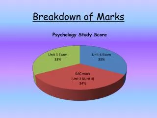
Breakdown of Marks. Exam Revision for Brain and Nervous System – Area of Study 1. The Brain. Cerebral Cortex Comprises two hemispheres (left & right) of nerve tissue Hemispheres are joined by a band of nerves called the corpus callosum which allows the hemispheres to communicate

E N D
Exam Revision for Brain and Nervous System – Area of Study 1
The Brain • Cerebral Cortex • Comprises two hemispheres (left & right) of nerve tissue • Hemispheres are joined by a band of nerves called the corpus callosum which allows the hemispheres to communicate • Each hemisphere has four lobes: frontal, parietal, occipital, temporal • Each hemisphere's surface is covered by a thin (3-5mm), convoluted (folded) layer of neurons called the cerebral cortex
Generally involved with information processing (e.g. perception, memory, thinking, language) • Neurons in the cerebral cortex process sensory and motor information
Frontal Lobe • Enables higher mental functioning involving learning and memory and control over complex movements, including the mechanics of speech (Broca’s area). • This region also controls emotions, personality and intellectual tasks requiring reasoning and planning.
Primary Motor Cortex • Controls voluntary movements of skeletal muscles • Motor cortex in left frontal lobe controls voluntary movements on right side of body and vice versa • Areas at top of motor cortex control lower parts of the body and vice versa • Amount of motor cortex is related to precision of movement of area (e.g. hands fingers, mouth, lips, have proportionately more motor cortex)
Broca’s Area • In left hemisphere only and involved in production of speech
Parietal Lobe • Mediates attention and is involved in spatial perception and bodily awareness based on sensations of touch pressure, pain and body movement.
Primary Somatosensory Cortex • Receives and processes sensory inputs from the body (i.e. body senses) • Sensory cortex in left parietal lobe processes sensory information from the right side of body and vice versa. • Amount of sensory cortex is related to sensory sensitivity of body area (e.g. mouth, lips, tongue and genital areas have proportionately more sensory cortex)
Occipital Lobe • The primary visual cortex enabling perception
Primary Visual Cortex • Receives and processes visual signals from two eyes • Right occipital lobe processes left visual field information (inputs from the right half of each eye) • Left occipital lobe processes right visual field information (inputs from the left half of each eye)
Temporal Lobe • The main location where auditory information is projected, enabling hearing to register. • This region left hemisphere of the brain contains Wernicke’s area, a language centre where speaking reading, writing and spelling are processed. • Also, this location acts as a centre for the recognition of different visual forms, enabling individuals to distinguish one object from another (role in memory)
Primary Auditory Cortex • Receives and processes auditory information from the ears • Left temporal lobe processes verbal sounds (e.g. words) • Right temporal lobe processes non- verbal (e.g. music)
Wernicke’s Area • In left hemisphere only involved in comprehension of speech
Hemispheric Specialisation • The tendency for one hemisphere to be dominant (i.e. more specialised, exert greater control) in certain cognitive and behavioural functions.
Hemispheric Specialisation Research on People with intact Brains • Psychological studies • Of people with intact (undamaged) brains indicate specialisation of left hemisphere for verbal tasks and specialisation of right hemisphere for non verbal tasks; however there is some overlap in functions between hemispheres.
Evidence for Specialisation includes Results of neuro-imaging studies • EEG studies • Brainwaves activity greater in left hemisphere with verbal tasks; greater in right hemisphere with spatial tasks • Tachistoscope Studies • Left hemisphere identifies verbal stimuli (words) more quickly when briefly flashed to right visual field and right hemisphere identifies non-verbal, spatial stimuli (e.g. pictures of objects) more quickly when flashed to the left visual field
Research On people with Split Brains • Split Brain Studies • Ability to verbally name and identify words and objects flashed to the right visual field (i.e. sent to left hemisphere); but • Inability to verbalise words or name objects flashed to the right visual field (i.e. sent to left hemisphere); but • Inability to verbalise words or name objects flashed to left visual field (i.e. sent to the right hemisphere); patients can identify these objects by touch alone using their left hand (i.e. controlled by the right hemisphere) • Severed corpus callosum prevents hemispheric exchange of information.
So, say a "typical" (language in the LEFT hemisphere) split-brain patient is sitting down, looking straight ahead and is focusing on a dot in the middle of a screen. Then a picture of a spoon is flashed to the right of the dot. The visual information about the spoon crosses in the optic chiasm and ends up in the LEFT HEMISPHERE. When the person is asked what the picture was, the person has no problem identifying the spoon and says "Spoon." However, if the spoon had been flashed to the left of the dot (see the picture), then the visual information would have travelled to the RIGHT HEMISPHERE. Now if the person is asked what the picture was, the person will say that nothing was seen!! But, when this same person is asked to pick out an object using only the LEFT hand, this person will correctly pick out the spoon. This is because touch information from the left hand crosses over to the right hemisphere - the side that "saw" the spoon. However, if the person is again asked what the object is, even when it is in the person's hand, the person will NOT be able to say what it is because the right hemisphere cannot "talk." So, the right hemisphere is not stupid, it just has little ability for language - it is "non-verbal."
Wada test • When a hemisphere is anaesthetised, limbs on opposite side of body go limp; when the left hemisphere is anaesthetised, language is impaired.
Central Nervous System • The central nervous system (CNS) the section of the nervous system that includes the brain and the spinal cord. • The spinal cord forms part of the central nervous system, and connects the brain to other areas of the body.
The Peripheral Nervous System (PNS) • The peripheral nervous system links the central nervous system to other parts of the body and is divided into the somatic nervous system and the autonomic nervous system.
Somatic nervous system in the control of skeletal muscles • The somatic nervous system transports messages to and from the sense organs and the spinal cord. • The somatic system, via its connections with the skeletal muscles, controls voluntary actions like waving one’s hand or other bodily movements. • Nerves from the somatic system do not control any of the non-skeletal muscles, such as the heart, lungs, stomach, intestines, etc.
Autonomic nervous system (ANS) in the control of the non skeletal muscles • The autonomic nervous system controls involuntary bodily activities, such as digestion and heart rate, when nerves transmit information to and from the glands and internal organs enabling a balanced state. Autonomic = Automatic The Autonomic Nervous System involves automatic (involuntary) processes.
Distinction between sensory and motor neuron activity within the somatic nervous system • Sensory (afferent) nerve fibres receive sensory stimuli arising inside or outside the body and transmit them inwards to the central nervous system where they are correlated. • The motor (efferent) nerve fibres transmit information outwards, away from the central nervous system, enabling the co-ordination of organs such as muscles and glands. • This allows them to work harmoniously ensuring the well being of the individual
Nervous System Sympathetic Increases responsiveness of muscles and organs during activity, stress or when threatened (e.g. fight or flight) Parasympathetic Decreases responsiveness of muscles and organs, thereby restoring the body’s functioning (e.g. relaxation response)
Imagining a parachute floating down to the ground may help you to remember the prefix for the Parasympathetic branch which brings physical mechanisms back down to normal levels again, relaxing the body after arousal.