Download
1 / 55
550 likes | 700 Views
呼吸系统基本病变的 CT 表现 THE BASIC PATHOLOGICAL CHANGES OF THE RESPIRATORY SYSTEM ON CT. 刘 源 (liu yuan) Department of radiology 1 st affiliated hospital Shantou university medical college. 一、 概述 (SUMMARIZE) 1. 基本病变与基本征象的关系

E N D
呼吸系统基本病变的CT表现 THE BASIC PATHOLOGICAL CHANGES OF THE RESPIRATORY SYSTEM ON CT 刘 源(liu yuan) Department of radiology 1st affiliated hospital Shantou university medical college
一、 概述(SUMMARIZE) 1. 基本病变与基本征象的关系 The relationship(connection) between basic pathological changes and basic sign 2. 基本征象在影像分析、诊断中的重要性 The significance of basic sign for imaging analysis and diagnosis 3. 与影像基本病变密切相关的组织学
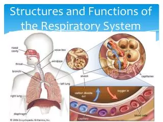
Secondary lobule Terminal bronchiole Respiratory bronchiole Alveolar duct Alveolar sac Pulmonaryalveoli 肺实质(lung parenchyma)
肺间质(interstitial tissue): 肺的框架结构-- 各级支气管和血管周围、小叶间隔、胸膜下和肺泡粘膜下的结缔组织
二、肺的基本病变 The basic pathological changes of lung • 渗出与实变(Exudation and consolidation) • 肺泡充实性病变 分肺实变(密度高于血管)和磨沙玻璃样影(密度低于血管) • 定义及病理(definition and pathology) • 常见疾病 炎症、肺水肿、肺泡蛋白沉着症 • CT表现(CT representation) • 形态(shape) 小片、大片、肺段性、大叶性、 弥漫性。 • 密度(density) 致密增高 支气管气相 • 层—层、肺窗—纵膈窗间的变化 • (changes between slices and windows)
2. 增殖性病变(proliferative lesion) • 定义及病理(definition and pathology) • 常见疾病 肉芽肿、炎性假瘤、慢性炎症 • CT表现(CT representation) • 形态 结节 、肿块 、大片状。小结节多为 肉芽肿,较大结节及肿块可为炎性假瘤,肿块性及肺段或肺叶实变影像可为慢性肺炎 • 边缘 清楚 • 密度(density) • 随访复查(follow up) 动态变化慢
3. 纤维性病变(fibrotic lesion) • 定义及病理(definition and pathology) • 常见疾病(main disease) • CT表现(CT representation) • 大小、形态、分布(size,shape,distribution) • 边缘(edge or margin) • 密度(density)
4. 钙化病变(calcification) • 定义及病理(definition and pathology) • 常见疾病 • 局限性:斑片块状:结核 爆花生米:错构瘤 • 弥漫性:细点状:肺泡微石症 小结节状:尘肺 • CT表现(CT representation) • 大小及形态(size and shape) • 边缘(edge or margin) • 密度(density) 一般CT值>100HU
错构瘤钙化 肺结核球中的钙化
5. 肿块和结节(pulmonary mass and nodule) • 定义及病理(definition and pathology) • 常见疾病(main disease) • CT表现(CT representation) • 肿块的确定(confirmation of the mass) • A.形态相对规则、轮廓较清,各径线接近 • B.密度相对较均匀 • C.层—层、肺窗—纵膈窗间变化小 • (相对于斑片状实变病灶)
具体表现(particular representation) • 部位(position) • 数目和大小(number and size) • 轮廓、形态(outline or shape) • 边缘(edge or margin) • 密度、内部结构(density,inside structure ) • 肿块强化(enhancement of the mass) • 邻近结构改变(changes around the mass ) • 随访复查(follow up)
肺门肿块 肿块强化
5 . 空洞(cavity) • 定义及病理(definition and pathology) • 常见疾病(main disease) • 空洞CT表现(CT representation of cavity) • 部位、数目和大小( position ,number and size) • 洞壁厚度(the wall thickness of cavity) • 虫蚀样空洞(moth-eaten cavity) • 薄壁空洞(thin-walled cavity) • 厚壁空洞(thick-walled cavity) • 洞壁外缘(outer edge of cavity, • as Exudation and consolidation, mass) • 洞壁内缘(inner edge of cavity, nodule of the wall) • 洞内气液平(air-fluid level) • 邻近结构改变(changes around the mass ) • 随访复查(follow up)
空腔(intrapulmonary air containing space ) • 定义及病理(definition and pathology) • CT的鉴别要点(main point of differentiation) • 囊腔壁(thickness and edge of the wall ) <1mm • 洞内气液平(air-fluid level) • 邻近结构改变(adjacent change )
1、小叶中心型 2、全小叶型 3、间隔旁或胸膜下 4、瘢痕旁型 早期肺气肿示意图
6. 肺不张(atelectasis) • 定义及病理(definition and pathology) • 分类(原因,程度)(type: cause,extent and degree) • CT表现(CT representation) • 密度(density) • 大小及形态(size and shape) • 边缘(edge or margin) • 邻近结构改变(changes around) • 与实变、肿块的鉴别