Download
1 / 1
10 likes | 136 Views
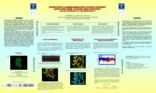
INTERACTION OF A BROMODOMAIN WITH A PEPTIDE CONTAINING ACETYLATED LYSINE: A DYNAMIC SIMULATION STUDY (Towards Identification of a “specificity domain”). X. Périole, E. L. Mehler and H. Weinstein Dept. of Physiology and Biophysics, Mount Sinai School of Medicine

E N D
INTERACTION OF A BROMODOMAIN WITH A PEPTIDE CONTAINING ACETYLATED LYSINE: A DYNAMIC SIMULATION STUDY • (Towards Identification of a “specificity domain”) X. Périole, E. L. Mehler and H. Weinstein Dept. of Physiology and Biophysics, Mount Sinai School of Medicine One G. Levy Place, New York, NY 10029. Conclusion Introduction The bromodomain (BRMD) is a highly conserved ~110 residues motif which forms a four-helix bundle1. It is usually contained in large multiprotein complexes involved in the transcription machinery, e.g. histone acetyltransferase (HAT)2. Although the specific function of the BRMD in HAT activity and/or in chromatin targeting and/or remodeling is still undefined, it is well known that it involves specific binding of acetylated-Lysine (AcK). Understanding the interaction of BRMDs with AcK-containing peptides will further our knowledge of how they contribute to transcription mechanisms. We report a study of structural and dynamic details of the interaction between the BRMD of the HAT co-activator P/CAF (p300/CBP-associated factor) and an 11 residue AcK-containing peptide (Fig.1). This study is based on structural data from NMR structure elucidation in the laboratoty of Ming-Ming Zhou at Mount Sinai School of Medicine. Computer simulations starting from this structure were carried out with a combination of Molecular Dynamics with Explicit and Implicit solvent models. Two systems were studied : 1) the wild type (WT) of the complex and 2) a mutant of the BRMD interacting with the same peptide. The purpose of the simulation of the WT is to study the dynamic properties of the BRMD-peptide interaction and the role of the solvent (water). The mutant was designed to explore the elements of specificity of the interaction with the peptide. The simulations are not completed, as the system had not yet reached equilibrium after 500 ps and 800 ps for the WT and V763G, respectively. Nevertheless, VAL763 is clearly emerging as part of the « specificity domain » in the interaction of the P/CAF BRMD with the peptide, for the following reasons : 1. the P/CAF and scGCN5 present different binding modes of a AcK-containing peptide around the position of the VAL763 in the P/CAF. 2. the close contact analysis of the NMR structures shows VAL763 as an important contact between the BRMD and the peptide but not participating in the interaction with the AcK. 3. the evolution of the distance between the C of the residue 763 (VAL and GLY for the WT and the mutant, respectively) and the C of the TYR02 of the peptide during the simulation, clearly indicates that the mutation decreases of the stability of the complex. 4. the difference between the energy averages we extract from the simulation, using an implicit solvent model (SCP-ISM 6, 7) favors the wild type. Longer simulations of the WT and V763G to reach final equilibrium are in progress. M E T H O D S M E T H O D S MOLECULAR DYNAMICS EXPLICIT SOLVENT IMPLICIT SOVENT We use a 48 Å sphere comprising ca. 14500 TIP3p water molecules that solvates any atom of the complex by at least 13 Å of free water molecules. The 3 Å outer shell of water molecules is subjected to harmonic constrains following the restrained water droplet model6. This allows the conservation of pressure and density conditions and is more economical than the typical Periodic Boundary Conditions approach. We use the new SCP-ISM3, 4 (Screened Coulomb Potential-Implicit Solvent Model). This model introduces: 1) a dielectric function of sigmoidal form to screen the Coulombic interactions, 2) a self-energy of the atoms calculated from the integral form of the Born equation and a novel approach to estimate Born radii of atoms in the protein environment3 and, 3) an approach4 to account for H-bond interaction based on the degree of exposure of the polar hydrogens to the proton acceptor environment. The method is parametrized in the context of the all-atom param22 force field. We use the CHARMM5 program package to study the dynamic of the complex. The starting structures of the simulations are extracted from a NMR model of the WT relaxed for 200ps with constrains. After that the WT and the mutant V763G are simulated free. The program was used with usual conditions: time step of 1 fs, SHAKE algorithm to constrain the H-bonds, and a 12 Å cutoff coupled to a shift function, ... The systems are thermalized to 310 K and equilibrated before the production period. Role of the Solvent in the BRMD-AcK Binding: Current Status of the Evolution of BRMD-peptide Binding During the Simulations Analysis of the NMR Models : A first analysis of 20 NMR models of the complex provided a list of residues from the BRMD involved in close contacts (<5Å) with the peptide in all the models : Table 1. Two residues are considered in contact in a NMR model if they have at least two atoms at a distance of < 5Å. From this list of contact residues a « specificity mutation » was chosen based on tree criteria : a) it shoud be involved in a close contact with the peptide, b) but not with the AcK, and c) it should not be a conserved locus in the different BRMDs (cf. Fig.2). In the Fig. 3 it appears that the peptide does not fill entirely the the binding cleft in the BRMD, above the AcK. Therefore, we considered the possible role of the water in supporting the interaction. Fig. 4 shows a number of water molecules in the cleft depicted during the 350-400 ps time frame in the simulation. This is comparable to the solvent distribution in the cleft of the scGCN5 BRMD-AcK peptide complex obtained from crystallography7, see Fig. 5. Although neither wild type or mutant simulations have reached their final equilibrium we present here the current status of the calculations. The comparative evolution of the structures reveals mechanistic differences between the complexes of the WT and V763G. A plot of the distances between the C of position 763 in the wild type and V763G mutant and the C of TYR02 of the peptide is shown in Graph 1. Graph 1: Distance between C s of VAL763 (WT) and the C of TYR02pept. The System : Figure 4 : positions of water (red dots) in the cleft during a 350-400 ps time frame Table 1 : The BRMD-peptide contacts. Figure 1: Acknowledgements. We thank M.-M. Zhou and L. Zeng for giving us access to the NMR structure models of the complex P/CAF-peptide before publication. R E S U L T S R E S U L T S Distances (A) References. 1. C. Dhalluin, et al., Nature399, 491 (1999). 2. F. Jeanmougin, et al. Trends Biochem. Sci. 22, 151 (1997) 3. S. A. Hassan et al., J. Phys. Chem.104, 6478 (2000) 4. S. A. Hassan et al., J. Phys. Chem.104, 6490 (2000) 5. B. R. Brooks et al., J. Comp. Chem.4, 187 (1983) 6. R. Sankararamakrishnan et al., Int. J. Quant. Chem.77, 174 (2000) 7. D. J. Owen, et al., EMBO J.19, 6141 (2000) The BRMD residues involved in a conserved contact with the peptide are : VAL752, GLU756, ALA757, PRO758, TYR760, VAL763, TYR802, TYR809 Times (ps) Figure 3 :The figure shows the different residues of the BRMD in contact with the peptide. The color code in the same as in the table. During the first 500 ps of the mutant simulation the C distance shortens, indicating that the Y02 which was interacting with VAL763 moves in to fill the space freed up by the mutation. In the last 300 ps, however, the distance increases due to the weaker interaction of the peptide with the mutant BRMD. The distance between the two C fluctuates more in the mutant complex than in the WT. This is illustrated by the standard deviations of these distances calculated between 186 ps to 486 ps : 0.46 and 0.70 Å respectively for the WT and V763G. This is representative of the fundamental difference between the two systems as the WT is much more stable than the mutant. Figure 5 : crystallographic positions of water (red dots) in the cleft The Bromodomain : 114 residues arranged in a four-helix bundle The peptide : 11 residues SER TYR GLY ARG AcLYS LYS ARG ARG GLN ARG CYS We evaluated the interaction energy of the BRMD with the peptide. Because the solvent has a crucial role in the interaction we used the SCP-ISM in the calculation of energies. The interaction energy, Eint, is define as : Figure 2:Alignment of Bromodomains1 The selected candidate for the specificity mutation is VAL763. The structure of the complex indicates that VAL763 is involved in a directed BRMD-peptide hydrophobic interaction and not with the AcK05. In addition this residue is not well conserved in the BRMDs, cf. Fig. 2. Note that in sgGCN5, there is a PHE in this position and the crystal structure revealed a different binding mode between the BRMD and a peptide7. To determine the role in the stabilization of the complex, VAL763 was mutated to GLY in the model. Eint=Etot-EBRMD-Epept WT: V763G: <Eint>186-486ps= -43.054.3 kcal/mole -40.836.7 kcal/mole