Download
1 / 54
660 likes | 1.25k Views
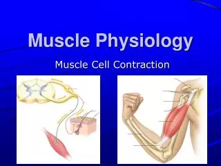
Muscle Physiology: The Actions of the Sarcomere. Cardiac Muscle Characteristics Intercalated disks Striated Involuntary Located in heart. Skeletal Muscle. Characteristics Many nuclei per cell Striated Voluntary Located along bones. Smooth Muscle. Nonstriated Involuntary

E N D
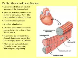
Cardiac Muscle • Characteristics • Intercalated disks • Striated • Involuntary • Located in heart
Skeletal Muscle • Characteristics • Many nuclei per cell • Striated • Voluntary • Located along bones
Smooth Muscle • Nonstriated • Involuntary • Located in digestive tract
Functions of Muscles • Movement: results from muscle contraction, enables you to respond quickly • Maintains Posture and Joint Stability: allows you to sit upright; stabilize joints of the body • Support Soft Tissue: abdominal muscles protect underlying digestive organs. • Guard Entrances and Exits • Generate Heat: heat is generated as they work…FRICTION • Maintains body temperature • Skeletal muscles create the most heat
Characteristics of Muscle Tissue • Excitability: ability to receive and respond to stimuli… • Contractibility:ability to shorten quickly and with force… • Extensibility: ability to be stretched or extended beyond their resting state… • Elasticity: ability of a muscle fiber to recoil and resume its resting length
Organization of Muscle • Muscles are composed of groups of fibers called fasicles. • Fibers are the muscle cells inside all muscle. • Tendons are bands of collagen fiber that attach muscle to bone.
Z line I band H band A band
Cross sectional view of Sarcomere. Differences are detected in the sizes of the myofilaments Myosin is the thicker fiber. Actin is the thinner fiber.
Sarcolemma Mitochondrion Myofibril Dark A band Light I band Nucleus (b) Diagram of part of a muscle fiber showing the myofibrils. Onemyofibril is extended afrom the cut end of the fiber. Striations are seen because of sarcomere bands.
Muscle fiber structure • Muscle cell • Sarcolemma • Sarcoplasm • Sarcoplasmic reticulum • T tubule • mitochondria
Sliding Filament TheoryActin slides over myosin shortening the sacromere between the Z lines
Events at the Neuromuscular Junction 1 Action potential arrives at axon terminal of motor neuron. Ca2+ Ca2+ 2 Voltage-gated Ca2+ channels open and Ca2+ enters the axon terminal. Synaptic vesicle containing ACh Mitochondrion Axon terminal of motor neuron Synaptic cleft 3 Ca2+ entry causes some synaptic vesicles to release their contents (acetylcholine) by exocytosis. Fusing synaptic vesicles ACh Junctional folds of sarcolemma 4 Acetylcholine, a neurotransmitter, diffuses across the synaptic cleft and binds to receptors in the sarcolemma. Sarcoplasm of muscle fiber
Myelinated axon of motor neuron Action potential (AP) Axon terminal of neuromuscular junction Nucleus Sarcolemma of the muscle fiber 1 Action potential arrives at axon terminal of motor neuron. Ca2+ Synaptic vesicle containing ACh Ca2+ 2 Voltage-gated Ca2+ channels open and Ca2+ enters the axon terminal. Mitochondrion Synaptic cleft Axon terminal of motor neuron 3 Ca2+ entry causes some synaptic vesicles to release their contents (acetylcholine) by exocytosis. Fusing synaptic vesicles Junctional folds of sarcolemma ACh 4 Acetylcholine, a neurotransmitter, diffuses across the synaptic cleft and binds to receptors in the sarcolemma. Sarcoplasm of muscle fiber Postsynaptic membrane ion channel opens; ions pass. 5 ACh binding opens ion channels that allow simultaneous passage of Na+ into the muscle fiber and K+ out of the muscle fiber. K+ Na+ Degraded ACh 6 ACh effects are terminated by its enzymatic breakdown in the synaptic cleft by acetylcholinesterase. Ach– Postsynaptic membrane ion channel closed; ions cannot pass. Na+ Acetyl- cholinesterase K+ Events at the Neuromuscular Junction
Setting the stage Axon terminal of motor neuron Action potential Synaptic cleft is generated ACh Sarcolemma Terminal cisterna of SR Ca2+ Muscle fiber Triad One sarcomere
Actin Troponin Tropomyosin blocking active sites Ca2+ Myosin Calcium binds to troponin and removes the blocking action of tropomyosin. 3 Active sites exposed and ready for myosin binding Contraction begins 4 Myosin cross bridge The aftermath
ADP P i 2 The power (working) stroke. Cross Bridge Cycle (2 of 4)
ATP 3 Cross bridge detachment. Cross Bridge Cycle (3 of 4)
ADP ATP hydrolysis PI 4 Cocking of myosin head. Cross Bridge Cycle (4 of 4)
Players for the power stroke • Cross bridge attachment • Power strokes • Cross bridge detachment • “Cocking” of the myosin head
Motor Unit: A motor neuron and all the muscle fibers it stimulates. Atrophy- when muscle fibers become weaker and smaller due to lack of stimulation by a motor neuron.
Muscle Tension • The amount of tension produced by a muscle is determined by: • The frequency of muscle stimulation. • The number of muscle fibers activated. • 3. Degree of stretch by sarcomere. (length-tension relationship • Myogram – a graph that measures tension developing in a muscle fiber.
Muscle that reaches peak tension during rapid cycles of contraction and relaxation.
Complete tetanus = relaxation state is eliminated. Recruitment –multiple motor unit summation
Stimulus strength Maximal stimulus Threshold stimulus Proportion of motor units excited Strength of muscle contraction Maximal contraction Relationship between stimulus intensity and muscle tension.
Muscle Metabolism • Muscle stores limited reserves of ATP ~ 4-6 Seconds • 3 Pathways for Generating ATP • Production of ATP from Creatine phosphate • Aerobic Respiration • Anaerobic Respiration
Aerobic Muscle Metabolism • Glycolysis • Aerobic Respiration • Krebs Cycle • ETC
Anaerobic Muscle Metabolism • Oxygen Debt • Lactic Acid Fermentation • Muscle Fatigue
Creatine Phosphate Muscle cells store 2-3 times creatine as ATP. Stored ATP and CP provide for maximum muscle power for 14-16s. (100 m dash) CP + ADP creatine kinase Creatine + ATP
(a) Direct phosphorylation (b) Anaerobic pathway (c) Aerobic pathway Coupled reaction of creatine Glycolysis and lactic acid formation Aerobic cellular respiration phosphate (CP) and ADP Energy source: CP Energy source: glucose Energy source: glucose; pyruvic acid; free fatty acids from adipose tissue; amino acids from protein catabolism Glucose (from glycogen breakdown or delivered from blood) Glucose (from glycogen breakdown or delivered from blood) CP ADP Creatine kinase O2 Glycolysis in cytosol Pyruvic acid Fatty acids Creatine ATP O 2 O2 Aerobic respiration in mitochondria Aerobic respiration ATP 2 Amino acids in mitochondria Pyruvic acid net gain O2 32 ATP CO2 Released to blood Lactic acid H2O net gain per glucose Oxygen use: None Oxygen use: None Oxygen use: Required Products: 1 ATP per CP, creatine Products: 2 ATP per glucose, lactic acid Products: 32 ATP per glucose, CO2, H2O Duration of energy provision: 15 seconds Duration of energy provision: 60 seconds, or slightly more Duration of energy provision: Hours 3 Pathways for regenerating ATP during muscle activity.
Prolonged-duration exercise Short-duration exercise ATP stored in muscles is used first. ATP is formed from creatine Phosphate and ADP. Glycogen stored in muscles is broken down to glucose, which is oxidized to generate ATP. ATP is generated by breakdown of several nutrient energy fuels by aerobic pathway. This pathway uses oxygen released from myoglobin or delivered in the blood by hemoglobin. When it ends, the oxygen deficit is paid back. Comparison of energy sources between short term exercise and prolonged exercise.
Isotonic and Isometric Exercise • Isotonic – tension increases and the muscle shortens • Lifting weights • Isometric – muscle does not shorten, the tension produced never exceeds resistanc • Trying to pick up a car
Red (slow) twitch fibers • Aerobic • Slow-acting ATPases (enzymes that break down ATP) • Large amounts of myoglobin • Red color to cell • Abundant supply of mitochondria • Fatigue resistant-as long as O2 is available • High endurance (jogging, swimming, soccer)
White (fast) twitch fibers • Large pale cells with twice the diameter of red fibers • Very little myoglobin • Contain fast-acting ATPases and contract rapidly • Contain few mitochondria, but large glycogen stores • Depend on anaerobic resp. to make ATP, therefore fatigues easily • Low endurance, much power….sprints