Download
1 / 31
310 likes | 582 Views
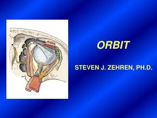
ORBIT R. Shane Tubbs, MS, PA-C, PH.D. BONY ORBIT. Supraorbital notch (foramen). Posterior ethmoidal foramen. Frontal bone. Anterior ethmoidal foramen. Orbital plate of ethmoid bone (lamina papyracea). Lesser wing of sphenoid. Lacrimal bone. Superior orbital fissure.

E N D
ORBIT R. Shane Tubbs, MS, PA-C, PH.D.
Supraorbital notch (foramen) Posterior ethmoidal foramen Frontal bone Anterior ethmoidal foramen Orbital plate of ethmoid bone (lamina papyracea) Lesser wing of sphenoid Lacrimal bone Superior orbital fissure Fossa of lacrimal sac Optic canal Orbital process of palatine bone Greater wing of sphenoid Zygomatic bone Inferior orbital fissure Maxillary bone Infraorbital groove & foramen
Brain Frontal sinus Ethmoidal cells Maxillary sinus
Ethmoidal cells Nasal septum Temporalis m. (in temporal fossa) Brain (in middle cranial fossa) Optic n. Sphenoidal sinuses Medial wall of orbit
SHEATHS OF THE OPTIC NERVE Central a. & v. of retina Pia Arachnoid Dura Subarachnoid space (Intervaginal space)
INTRINSIC MUSCLES OF EYEBALL Cornea Sclera Iris folds Lens Ciliary m. (accommodation) Dilator m. of pupil (mydriasis) Fibers of ciliary zonule (suspensory lig. of lens) Ciliary body Sphincter m. of pupil (miosis)
MUSCLES OF THE UPPER EYELID Levator palpebrae superioris Superior tarsal muscle
Superior oblique m. Levator palpebrae superioris m. Trochlea (pulley) Superior rectus m. Medial rectus m. Common annular tendon Lateral rectus m. (cut) Inferior rectus m. Inferior oblique m.
Superior tarsal plate Trochlea Levator palpebrae superioris m. Superior oblique m. Superior rectus m. (cut) Medial rectus m. Lateral rectus muscle Inferior rectus m. Superior rectus m. (cut) Levator palpebrae superioris m. (cut)
MOVEMENTS AROUND THE VERTICAL AXIS Inferior oblique Superior oblique Medial rectus Superior rectus Lateral rectus Inferior rectus ABDUCTORS ADDUCTORS
MOVEMENTS AROUND THE LATEROMEDIAL (TRANSVERSE) AXIS Inferior oblique Superior oblique Superior rectus Inferior rectus ELEVATORS DEPRESSORS
MOVEMENTS AROUND THE A-P AXIS Superior rectus Superior oblique INTORSION Inferior rectus Inferior oblique EXTORSION
Medial check ligament Lateral check ligament Periorbita Periorbita Medial rectus muscle and sheath Bulbar sheath (Tenon’s capsule) Lateral rectus muscle and sheath Orbital fat
L N F N. F. L.
POSITION OF CILIARY GANGLION Optic nerve Lateral rectus Ciliary ganglion Short ciliary nerves
Medial palpebral a. Lateral palpebral a. Supratrochlear a. Dorsal nasal a. Supraorbital a. Anterior ethmoidal a. Posterior ciliary aa. Posterior ethmoidal a. Continuation of ophthalmic a. Lacrimal a. Muscular branch Central a. of retina Ophthalmic a. Internal carotid a.
Supratrochlear v. Supraorbital v. Superior ophthalmic v. Angular v. Cavernous sinus Vorticose vv. Facial v. Inferior ophthalmic v. Pterygoid plexus