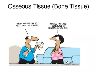
Tissue Integrity
Tissue Integrity . Susan Fowler sfowler@tricountycc.edu. Objectives p. 4. The concept of tissue integrity will be discussed as well as tissue damage. Tissue damage will be demonstrated by the exemplars of pressure ulcers and wound care. Tissue Integrity.

Tissue Integrity
E N D
Presentation Transcript
Tissue Integrity Susan Fowler sfowler@tricountycc.edu
Objectives p. 4 • The concept of tissue integrity will be discussed as well as tissue damage. • Tissue damage will be demonstrated by the exemplars of pressure ulcers and wound care.
Tissue Integrity • Defined by intact skin and mucous membranes with no evidence of damaged or destroyed tissue. • When tissue is damaged or destroyed, it is said to have lost its integrity. • Even when there is no damage evident, there may be a risk d/t factors present in the client’s internal or external environment.
Risk Factors for Tissue Damage • Wide variety of disease processes (mental and physical) • Nutritional state • Age • Immobility • Moisture • Shear and friction • Written as “Risk for Altered Tissue (or Skin) Integrity
Actual Tissue Damage • May be surgical wound or trauma • Surgical wounds can be open or closed • Traumatic wounds can be intentionally inflicted or unintentionally inflicted. • Risk factors coupled with neglect can cause a kind of tissue damage called pressure ulcers.
Pressure Ulcer • A localized area of tissue necrosis caused by unrelieved pressure that occludes blood flow to tissues. • Usually located over body prominences • Most common sites are sacrum and heels
Influencing Factors • Amount of pressure • Length of time pressure is exerted • Ability of tissue to tolerate externally applied pressure
Contributing Factors • Shearing force—pressure exerted on the skin when it adheres to the bed and the skin layers slide in the direction of body movement. • Friction—two surfaces rubbing against each other • Excessive moisture
Clinical Manifestations • Ulcers are graded or staged according to the deepest level of damage • Stage I (minor) to Stage IV (severe) p. 1283 • Slough or eschar may need to be removed to accurately stage some ulcers • Ulcers also may be classified as red, yellow, or black to help determine the best tx. p. 1285
Stage I • Persistent redness in lightly pigmented skin • Red, blue, or purple in darker skin • May be warm to touch • May have poor sensation • Does not blanch • Skin may be boggy, swollen, or thin over site, but no skin break is evident
Stage II • Skin is broken with partial thickness loss of epidermis, dermis or both • Presents as an abrasion, skin tear, intact or ruptured blister, or shallow crater
Stage III • Full-thickness loss involving damage or necrosis of subcutaneous tissue that may extend down to, but not through, underlying fascia • Presents as a deep crater with possible undermining of adjacent tissue
Stage IV • Full-thickness loss can extend to muscle, bone, or supporting structures • Bone, tendon, or muscle may be visible or palpable • Undermining and sinus tracts may also exist
Complications • Recurrence is most common • Infection—fever, leukocytosis, pain, increase in size, odor, or drainage • Cellulitis—surrounding tissue infection • Chronic infection—may persist for months • Septicemia (blood infection) • Osteomyelitis (bone infection)
Assessment • On admission (RN) and periodically according to protocol • Use assessment tool such as Braden Scale • Use inspection and palpation to assess color, breakdown, and temperature • Use natural or halogen light rather than fluorescent to assess dark skin • Ask patient how it feels—is it painful, itchy, or numb?
Expected Outcomes • No deterioration • Reduce contributing factors • No presence of infection • Heal without complications or recurrence
Interventions: Prevention • Identify risk factors • Implement prevention strategies: • Remove excessive moisture • Good skin and incontinence care • Avoid massage over bony prominences • Turn every 1 to 2 hours and avoid shearing • Use lift sheets • Pillows, heel and elbow protectors • Specialty beds
Prevention cont’d • Caloric intake elevated to 30 to 35 cal/kg/d or 1.25 to 1.5 g protein/kg/day • Supplements, enteral (GI), or parenteral (IV) feedings may be necessary • Keep patients as mobile as possible
Interventions: Treatment • Document size, stage, location, exudate, infection, pain, and tissue appearance • Keep ulcer bed moist • Cleanse with nontoxic solutions (saline) • Debride by medications or refer for surgical debridement • Adhesive membrane, ointment, moisture-retentive dressings • Teach self-assessment and self-care
Interventions: Operative Repair • Skin grafts • Skin flaps • Musculocutaneous flaps • Free flaps • Surgical debridement
Interventions: Patient/Family Education • Assess resources • Explain risk factors and causes • Teach incontinence care • Demonstrate correct positioning, turning • Teach daily inspection • Teach wound care • Stress good nutrition
Evaluation • Prevention strategies implemented • Wound has not deteriorated • No complications • Wound healed with no recurrence • Patient/family understands instructions
Wound Healing • Primary—straight line with all layers well-approximated, free of infection, no separation, fast healing, minimal scarring • Secondary—healing from inside out by granulation, increased infection risk, slow healing, extensive scarring • Tertiary—delay of 3-5d between injury and suturing, increased chance of infection and separation
Healing Phases • Initial (3-5d)—approximation, epithelial cell migration, mesh and initial capillary growth • Granulation (5d-4wk)—fibroblast migration, collagen formation, capillary beds formed, fragile tissue • Scar contracture (7d-mos)—remodeling of collagen, strengthening of scar
Factors Affecting Healing • Age—younger heals quicker • Nutritional status—malnourished and obese • Systemic disorders—DM, circulatory probs, immunosuppression • Presence of foreign bodies • Infection • Meds—corticosteroids, antibiotics, anticoagulants
Factors cont’d • Irradiation • Treatment of wound • Wound stressors—coughing, straining, vomiting, trauma • Type of wound • Presence of drains • Race—keloid formation in darker races
Complications of Healing • Hemorrhage • Infection • Dehiscence and evisceration
Nursing Responsibilities R/T Wound Care • Goal is for wound to remain intact with no complications • Assessment-size, color, drainage p. 1287 • Dsg change may be simple or complex • Remove staples and apply steri-strips • Don’t forget patient education and documentation
Documentation of Wound Care • “Sterile dsg change performed on abd wound. Old 4x4 dressing clean and dry. Wound 10 cm, closed, edges well-approximated, staples intact, without redness or drainage. Cleaned with 4x4 and normal saline and sterile 4x4 applied. Pt tolerated without complaints.”
Documentation of Complicated Wound Care • “Patient premedicated with Lortab ii tabs po for dsg change. Sterile dsg change performed on abd wound after 30 minutes. Outer ABD pad with 4x3 cm area of serosanguineous drainage. Removed two 4x4s 50% saturated with serosanguineous drainage. Removed 12 cm of serosanguineous saturated NuGauze packing. Incision open, 8(L)x4(W)x3(D)cm. No signs of infection present….”
Documentation cont’d • Granulation noted in wound bed. Incision irrigated with 60 mL sterile saline. Aerobic culture taken as ordered. Wound packed with 12 cm saline soaked NuGauze, covered with 2 sterile 4x4s and ABD pad. JP drain emptied of 45 mL of serosanguineous drainage. No odor or clots present. Pt tolerated with minimal discomfort. Culture taken to lab.”