Download
1 / 59
590 likes | 991 Views
Genetic Mutations. The following slides explain the molecular biology behind genetics cases included with the Case It v5.03 download. Online descriptions of these cases can be found at http://caseit.uwrf.edu ('access cases' link). Question #1.

E N D
Genetic Mutations The following slides explain the molecular biology behind genetics cases included with the Case It v5.03 download. Online descriptions of these cases can be found at http://caseit.uwrf.edu ('access cases' link)
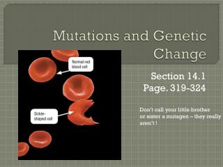
Question #1 REVIEW: What kind of mutation results from a change in a single DNA base? - a base substitution mutation What is an example of a disease caused by this type of mutation? - sickle-cell anemia
Sample case – sickle cell Case A: Steve and Martha are expecting their second child. They know that sickle cell anemia runs in both of their families. They want to know whether this child could be affected. Neither they nor their 10-year-old daughter, Sarah, have shown any symptoms of the disease. They decide to have DNA tests to determine the status of the fetus, as well as to find out whether they in fact are carriers of the disease gene.
Background – sickle cell Sickle cell anemia is a disease of red blood cells. It is caused by a mutation in the hemoglobin gene. A single base change results in a single amino acid substitution. This mutation causes the hemoglobin to change its conformation to a more elongated form under certain conditions, distorting the red blood cells and impairing their ability to carry oxygen. Sickle cell anemia is considered a recessive trait, since both chromosomes have to carry the mutation in order for the full blown disease symptoms to appear.
Background – sickle cell The sickle cell mutation also eliminates a restriction enzyme site - the recognition site for the enzyme MstII. To detect the sickle cell mutation, a patient’s DNA is digested with MstII and a Southern blot is performed using a probe corresponding to this region of the hemoglobin gene. The presence or absence of the sickle cell mutation can be determined based on the size of the fragment identified by the probe.
Question #2 REVIEW: What restriction enzyme site is eliminated by the sickle-cell mutation? - MstII - cctNagg is the recognition site (N can be any base) - abnormally long fragment results What techniques are used to detect the sickle-cell mutation? - RFLP with restriction enzyme MstII - (RFLP = Restriction Fragment Length Polymorphism) - Southern blotting with sickle-cell probe
Question #3 A mutation that causes the removal of base pairs from a DNA sequence is called what? - a deletion mutation
Question #3 continued Give an example of a human genetic condition caused by a deletion mutation. - cystic fibrosis is caused by deletion of 3 DNA bases - missing codon in mRNA - amino acid phenylalenine therefore missing from transmembrane protein in lungs
Sample case – cystic fibrosis Case B: (Contributed Stephanie Dahlby, Dan Tally, and Janelle Veerkamp, Biol 305 Students, Spring 1997, UW-River Falls) Lynda and Jim are expecting their first child. Recently, however, they learn that Lynda’s aunt died of CF and Jim’s uncle died of CF. They are worried that they might be carriers for the disease and pass cystic fibrosis on to their unborn child. They learn about a procedure which can determine whether they are carriers. They also learn about a procedure called amniocentesis which can detect if their unborn child has CF or is a carrier. However, amniocentesis is a very risky procedure. Jim and Lynda ultimately decide that they first want to be tested to see if they are carriers for the disease. If they learn that they both are carriers, they would like to go through with the amniocentesis to see if their child is affected.
Background – cystic fibrosis Background: Cystic fibrosis (CF) is generally considered the most common severe autosomal recessive disorder in the Caucasian population, with a disease frequency of 1 in 2,000 and a carrier frequency of 1 in 20. The major clinical symptoms include chronic pulmonary disease, pancreatic insufficiency, and an increase in sweat electrolyte concentrations. The cause of the disease appears to be a mutation in the gene encoding the cystic fibrosis transmembrane conductance regulator (CFTR), a membrane protein involved in transporting ions across epithelial surfaces, such as the linings of the lungs and intestines.
Background – cystic fibrosis Several mutations have been identified as being associated with a non-functional CFTR protein. The most common mutation, accounting for about 50% of CF cases, is called delta F508; it is a three-base deletion resulting in the loss of a phenylalanine at position 508, in the ATP-binding portion of the protein. This mutation is detected by sequence analysis of PCR-amplified DNA, or by hybridization with mutation-specific probes (the latter method is illustrated in Case B).
Question #4 What technique is used to detect the cystic fibrosis mutation in Case A of the Case It! exercise? - RFLP in region linked to mutated gene - loss of MspI site (ccgg) - PCR used to amplify linked region of DNA, then cut with MspI - Southern blotting not necessary since not dealing with lots of fragments
Question #4 continued What technique is used to detect the cystic fibrosis mutation in Case B of the Case It! exercise? - Southern blot with probe specific for mutation - no RFLP necessary (no restriction enzyme needed) - to determine genotypes, must run separately with normal probe and mutant probe, then compare blots
Question #5 A mutation caused by adding base pairs to a DNA molecule is called what? - an insertion mutation
Question #5 continued Give an example of a human genetic condition caused by an insertion mutation. - Huntington’s disease (chorea) is caused by the repeated insertion of the triplet CAG …...CAGCAGCAGCAGCAG…... - more than 50 repeats of the triplet causes disease - progressive degeneration of nervous system
Sample case – Huntington’s Case A: Susan is a 23-year-old whose father, age 55, and paternal aunt, age 61, have been diagnosed with Huntington’s chorea. A paternal uncle, age 66, appears to be unaffected by the disease. Susan wants to know if she inherited the mutated gene from her father so that she can prepare for that future if necessary. She arranges to undergo DNA testing for Huntington’s disease. Her 17-year old brother, John, also decides to be tested after talking with Susan.
Background – Huntington’s Huntington’s chorea is a neurodegenerative disease characterized by motor, cognitive, and emotional symptoms. The age of onset for symptoms is generally 30-50 years. The genetic basis of the disease is an amplification in a gene with an (as yet) unknown function. A triplet (CAG) is repeated 20-50 times in asymptomatic individuals; having more than 50 repeats is associated with disease symptoms.
Background – Huntington’s This amplification can be detected by restriction enzyme digestion and Southern blot analysis, since the size of the fragment bound by the probe is increased as a result of the amplification of the triplet repeat. Huntington’s disease is considered a dominant disorder, since one copy of the amplified gene appears to be sufficient to cause disease symptoms.
Question #6 What techniques are used to detect mutation for Huntington’s disease? - RFLP with EcoRI restriction enzyme - Southern blot with Huntington’s probe - mutated fragments are larger because of repeats
Question #7 What disease is caused by the deletion of one or more exons in a particular gene? - Duchenne’s muscular dystrophy (DMD) - characterized by progressive muscle weakness - a sex-linked characteristic - affects primarily males
Question #7 continued What are exons? - the parts of the DNA sequence that are actually used when a particular protein is made What are introns? - the parts of the DNA sequence not used (ignored) -the introns are cut out of the mRNA, then exons are spliced together to make functional mRNA
Sample case – DMD Case A: Jean and Bill have three sons, ages 12, 8, and 7, and a daughter, age 6. The oldest son and daughter are healthy, but the two younger sons are exhibiting symptoms of muscle weakness consistent with early muscular dystrophy. Jean knows that she has a family history of muscular dystrophy, but she does not know whether she is a carrier of the disease gene. She seeks DNA testing to determine whether her younger sons may have inherited the form of the dystrophin gene associated with Duchenne's muscular dystrophy (DMD).
Background - DMD One form of inherited muscular dystrophy, Duchenne’s, is X-linked and therefore affects primarily males. The symptoms of Duchenne's muscular dystrophy (DMD) include progressive and severe skeletal muscle weakness. A common mutation associated with DMD is a deletion of one or more exons in the dystrophin gene. These deletions can be detected by restriction enzyme digestion and Southern blotting using a combination of probes that will bind to multiple dystrophin exons.
Question #8 What technique is used to detect the DMD mutation? - RFLP with enzyme HindIII to separate exons - Southern blot with DMD probe “cocktail” - look for missing exons
Question #9 What are two mutations associated with Alzheimer’s disease? - mutation in codon 693: glutamic acid changed to glycine - loss of a recognition site for restriction enzyme MboII (gaaga)
Sample case – Alzheimer Case A: Martha, age 71, has been exhibiting increasingly severe symptoms of senile dementia and has been hospitalized for testing. She is in good health otherwise. Her three children - Sam (age 43), Joan (age 41) and Robert (age 38) - want to find out the cause of the dementia and determine the prognosis for Martha's future condition. They are also concerned that Martha may have a form of familial Alzheimer disease and want to know if they are at risk. The physician decides initially to test Martha for two mutations, 693 Gly and 717 Ile, in the amyloid precursor protein (APP) gene which are associated with inherited Alzheimer disease.
Background – Alzheimer Alzheimer disease is by far the most common cause of dementia in aging persons. The disease symptoms are identical to other forms of senile dementia, and diagnosis had been possible only at autopsy by the detection of protein clusters called amyloid plaques in the cerebrum. The disease is multifactorial and inheritance patterns are complex. Some forms of familial Alzheimer disease appear to be inherited as autosomal dominant traits, while others are recessive. Spontaneous Alzheimer disease also can occur in the absence of inherited factors.
Background – Alzheimer Mutations in at least four genes have been linked to Alzheimer disease. One of these is the amyloid precursor protein (APP) gene, which encodes the b-amyloid peptide found in the cerebral plaques of Alzheimer patients. The function of APP is not yet known, but certain APP point mutations are associated with inheritance of late-onset Alzheimer disease in some families. Two examples which can be detected by RFLP analysis are the codon 693 Glutamic acid to Glycine mutation and the codon 717 Valine to Isoleucine mutation. The 693 mutation results in the loss of a MboII site, while the 717 mutation results in the gain of a BclI site.
Question #9 - mutation in codon 717: valine changed to isoleucine - gain of a recognition site for the restriction enzyme BclI (tgatca) What technique is used to detect these mutations? - RFLP with MboII and BclI - Southern blotting with APP probe -look for abnormally large fragment (693 mutation) or abnormally small fragment (717 mutation)
Question #10 What are some mutations associated with breast cancer? -AG deletion (“185delAG” mutation) - TCAA deletion (“4184delTCAA” mutation) - C insertion (“5382insC” mutation) Technique used to detect mutations: - DNA amplifed by PCR (already done in Case It! example) - Southern blot using probe specific for mutation (no RFLP necessary) - if mutation present, 80% risk of breast cancer
Sample case – breast cancer While Elizabeth is reading the morning newspaper, she notices an ad for a free genetic screening for breast cancer at the clinic next week. The ad specifically invites women of Ashkenazi Jewish ancestry to participate. According to the newspaper ad, subjects will be tested to see whether they have mutations in the BRCA1 gene which would predispose them to breast cancer. Elizabeth, age 27, had heard about the discovery of the gene and about the mutation linked to Jewish women.
Sample case – breast cancer Her paternal grandmother had been diagnosed with breast cancer at age 51 and died two years later, and Elizabeth worried that she had inherited the disease. She also worried about her mother, age 52 and apparently cancer-free so far, and her 7-year old daughter. Her daughter is not allowed to participate in the screening, but Elizabeth convinces her mother to go with her to get tested.
Background – breast cancer Breast cancer is the most common malignancy among women. Current estimates are that one in eight women born in 1990 will contract breast cancer by age 85. Many factors contribute to breast cancer risk. Inheritance of breast cancer susceptibility genes contribute to approximately 5-10% of all breast cancers. The breast/ovarian cancer susceptibility gene BRCA1 has been identified on chromosome 17. Women who inherit certain BRCA1 mutations have an 80% risk of breast cancer.
Background – breast cancer BRCA1 appears to encode a tumor suppressor protein. Mutations that affect the function of this protein cause increased rates of cell division and a predisposition towards the development of malignancy. Several BRCA1 mutations, including point mutations, deletions, and insertions, have been identified that may contribute to loss of tumor suppressor function. These mutations can be identified by amplifying portions of the BRCA1 gene by PCR and then using RFLP analysis, direct sequencing, or hybridization with specific probes to detect the presence of mutations.
Background – breast cancer For the screening, a small amount of blood is drawn. DNA is isolated from the blood, and part of the BRCA1 gene is amplified by PCR. The amplified DNA is run on a dot blot with specific probes corresponding to mutations known to be linked to increased breast cancer susceptibility. The probe will only bind to the DNA if that mutation is present. DNA samples known to have specific mutation also are included. If a mutation is detected, use the probe corresponding to the normal sequence for that mutation site to determine whether the individual is homozygous or heterozygous for the mutation.
Question #11 What mutation is associated with phenylketonuria (PKU)? - C to T base-substitution mutation results in wrong amino acid (similar to sickle-cell mutation) - can’t metabolize amino acid phenylalanine - phenylalanine present in aspartame (artificial sweetener found in diet soft drinks, etc) - mental retardation and other problems