Download
1 / 23
230 likes | 324 Views
Understand the developing embryo phases, CNS protective mechanisms, hemispheric asymmetry, and prenatal brain development. Explore disorders associated with failures in protective systems and neuroanatomical features of conditions like fetal alcohol syndrome and autism. Gain insights into the functional neuroanatomy of the cerebellum and cranial nerves. Discover the impact of prenatal ethanol exposure on neurodevelopment. Explore autism neuropathology and diagnostic standards. Study hemispheric asymmetry using various techniques and imaging methods.

E N D
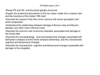
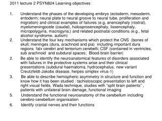
2011 lecture 2 PSYN824 Learning objectives • Understand the phases of the developing embryo (ectoderm, mesoderm, entoderm; neural plate to neural groove to neural tube, proliferation and migration) and clinical examples of failures (e.g, anencephaly (rostral), myelomeningocele (caudal), holosprosencephaly, lissencephaly, micropolygyria, macrogyria;) and related postnatal conditions (e.g., fetal alcohol syndrome, autism) • Understand the four key mechanisms which protect the CNS (bones of skull; meninges (dura, arachnoid and pia) including important dura regions falx cerebri and tentorium cerebelli; CSF (contained in ventricles, sub arachnoid and subdural spaces; Blood brain barrier) 2. Be able to identify the neuroanatomical features of disorders associated with failures in the protective systems arise and their clinical presentations (subdural haematoma, hydrocephalus, new variant Creutzfeldt-Jakobs disease, herpes simplex virus-1) 3. Be able to describe hemispheric asymmetry in structure and function and know how it has been studied : tachistoscopic presentation to left and right visual fields, Wada technique, studies with “split brain patients” , patients with unilateral brain damage, functional imaging 5. Understand the functional neuroanatomy of the cerebellum including cerebro-cerebellum organisation 6. Identify cranial nerves and their functions
Prenatal brain development • Three layered structure: • endoderm (digestive tract, respiratory system) • mesoderm (skeleton and muscles) • ectoderm (cns and skin) • Neurulation: neural groove becomes neural tube • Fusing of rostral (partial closure results in anencephaly) then caudal end (failure results in myelomeningocele) • To-be-neurons and glia proliferate, migrate and differentiate • Neuroanatomical presentation of • holoprosencephaly (complete or partial absence of the forebrain) • lissencephaly (“smooth brain”) • micropolygyria • macrogyria
adapted from Burd et al (2003) Fetal alcohol syndrome: neuropsychiatric phenomics. Child Neuropsychology, 25, 697-705 Prenatal ethanol exposure → decreased neuron production → abnormal migration [small brain] [structural abnormalities] → abnormal neurotransmitter levels → changes in electrical signalling → abnormal apoptosis → cns dysfunction → impairments in test performance
Astley et al (2009) Alcoholism: Clinical and Experimental Research,33(10), 1671–1689lFacial features of associated with Fetal alcohol syndrome
FAS Most consistent neuroanatomical abnormalities are • microcephaly • corpus callosum including complete or partial agenesis, volume reductions, particularly in the anterior and posterior sections • reductions in other white matters pathways esp in frontal lobes, and tracts connecting the occipital lobes with parietal and frontal lobes • cerebellum: decreased surface area and volume, particularly in anterior vermis • decreased volume of caudate nucleus (this may compromise fronto-striatal circuits
Astley et al (2009) Two subjects with (A) agenesis and (B) hypogenesis of the corpus callosum in the fetal alcohol syndrome (FAS) ⁄ partial FAS group. (C) Variability in corpus callosum shape among Controls with no prenatal alcoholexposure.
Autism • Originally described by Kanner in 1943 the core features (aka the autistic triad) • (a) impairments in reciprocal social interactions (b) an abnormal development and use of language; and (c) repetitive and ritualized behaviours, restricted range of interests • Co-morbid with developmental delay, epilepsy and anxiety and mood disorders • Diagnostic standards: no gold standard yet ? • Neuroanatomy: • Altered developmental time course in size of whole brain size, and probably frontal lobe, and amygdala • Smaller corpus callosum and cerebellar vermis, with fewer Purkinge cells
Neuropathology of autism Alexander et al (2007) NeuroImage 34, 61–73 Global and regional corpus callosum regions of interest for one subject with autism in (a) the mid-sagittal plane, (b) a coronal plane through the mid-body, and (c) the axial plane depicting regions of the genu and splenium. The total corpus callosum is shown in red, genu in yellow, body in purple and splenium in blue.
Hemispheric asymmetry in structure and function been studied using : • tachistoscopic presentation to left and right visual fields, • Wada technique, • studies with “split brain patients” http://www.youtube.com/watch?v=lfGwsAdS9Dc • patients with unilateral brain damage • functional imaging Diagram of visual pathway from Kapit & Elson (2003) Note • axons from the retina on the nasal side cross at the chiasm, those on the lateral side (K4 and J4) do not • Relay point in lateral geniculate nucleus of thalamus (K6 and J6) • Pathway to superior colliculi (K7 and J7) in the midbrain
From Paul et al (2007) Nature Neuroscience Reviews, 8, 291-299” prefrontal light green; pre-motor and suppl. motor light blue; motor dark blue; sensory red; parietal orange; occipital yellow, temporal violet;From Witelson (1998) Brain, 112, 799-835: 1 rostrum; 2 genu; 3 rostral body, 4 anterior mid body; 5 posterior mid body; 6 isthmus; 7 splenium; typically 3-5 called the body
left hemisphere sees chicken → right hand chooses chicken claw right hemisphere sees snow scene → left hand chooses shovel when asked why the shovel was chosen the speaking hemisphere says “ to clear out the chicken shed”
Cerebellum: External landmarks are the vermis, anterior, posterior and flocculonodular lobes; primary and horizontal fissures; tonsils; superior, middle and inferior cerebellar peduncles cortex highly structured with distinct cell types called molecular, purkinje cell and granule cells Internal landmarks: the arbor vitae and four pairs of irregularly shaped nuclei from medial to lateral they are Fastigial , Globose, Emboliform n and Dentate n (Don’t Eat Greasy Food) . The globose and emboliform are collectively called the interposed n. Reciprocal connections to the brainstemand cortex via the cerebellar peduncles, pons, red nucleus, various nuclei of the thalamus
cerebellum Two principles of organisation: • Unilateral motor signs after unilateral damage • crossed cerebro-cerebellar organisation (left hemisphere of cerebral cortex projects to the right cerebral hemisphere and vice a versa, shown by fMRI in normals, reorganisation after stroke and reorganisation after congenital left hemisphere damage) Vestibular signs (emesis, dizziness and vertigo) result from lesions of vermis in middle region of posterior lobe and in the flocculonodular lobe Motor signs: damage to vermis in anterior lobe and superior portions of posterior lobe cause oculomotor abnormalities, gait ataxia, appendicular ataxia and dysmetria, dysarthric speech and oculomotor abnormalities Cognitive signs: lateral regions of posterior lobe
Schmahmann’s (1998) proposal of cerebellar cognitive affective syndrome or dysmetria of thought Non-motor roles of the cerebellum: inferred from signs and symptoms after damage to the lateral portions of the posterior hemisphere and from fMRI studies disturbances of executive function: poor performance on tasks of planning, set shifting, abstract reasoning, verbal fluency and perseveration, naming and word stem completion, working memory, impaired spatial cognition: including visuospatial disorganisation and impaired visuo-spatial memory cerebellar mutism linguistic difficulties: dysprosodia and agrammatism personality change: blunting of affect, disinhibited or inappropriate behaviour Absence of phonological similarity effect: bead peace leaf tease deal vs bead pace ledge tab dip Vs bell keg ledge neck set vs feet lake deaf mass mud