Download
1 / 7
70 likes | 311 Views
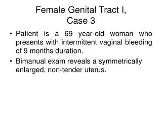
Female Genital Tract I, Case 3. P atient is a 69 year-old woman who presents with intermittent vaginal bleeding of 9 months duration. B imanual exam reveals a symmetrically enlarged, non-tender uterus. Identify the organ Describe the gross findings. Endometrial cavity. Tumor.

E N D
Female Genital Tract I,Case 3 • Patient is a 69 year-old woman who presents with intermittent vaginal bleeding of 9 months duration. • Bimanual exam reveals a symmetrically enlarged, non-tender uterus.
Identify the organ Describe the gross findings
Endometrial cavity Tumor Myometrium Uterus – endometrial cavity A diffuse tumor involves the entire endometrial surface
Identify the organ Identify the structures Describe the gross findings
A – Endometrial Cavity B – Endocervical Canal C - Ectocervix Endometrial Adenocarcinoma The surgical specimen consists of the entire uterus, right ovary, and fallopian tube. The uterus is opened along the anterior midline. A polypoid carcinoma (arrow) is present in the upper left corner of the endometrial cavity. Note that the remainder of the cavity is lined by flat, normal epithelium.
Endometrial Adenocarcinoma A close-up view of the endometrial cavity reveals a polypoid carcinoma (arrow). The mucosa lining the remainder of the cavity is normal. A = Myometrium
Endometrial biopsy - Describe the histologic findings Microscopic section of the endometrium reveals an adenocarcinoma. The neoplasm is composed of well differentiated glands which are "back to back". Very little stroma exists between the glands. The glands are lined by multilayered, atypical epithelium. Numerous mitoses (arrows) are present.