Download
1 / 30
330 likes | 526 Views
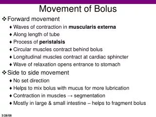
Movement of Bolus. Forward movement Waves of contraction in muscularis externa Along length of tube Process of peristalsis Circular muscles contract behind bolus Longitudinal muscles contract at cardiac sphincter Wave of relaxation opens entrance to stomach Side to side movement

E N D
Movement of Bolus • Forward movement • Waves of contraction in muscularis externa • Along length of tube • Process of peristalsis • Circular muscles contract behind bolus • Longitudinal muscles contract at cardiac sphincter • Wave of relaxation opens entrance to stomach • Side to side movement • No set direction • Helps to mix bolus with mucus for more lubrication • Contraction in muscles → segmentation • Mostly in large & small intestine – helps to fragment bolus
Digestive System Chapter 22 – Day 3
Stomach Anatomy • Shape • Sphincters • Cardiac • Pyloric • Folds = rugae • Deep muscular folds • Mucosa Fig. 22.12
Stomach Anatomy • Mucosa • Gastric pits with gastric glands • Secretory cells • 4 types of secretory cells: Cell Secretion • Chief cells → Pepsinogen • Parietal cells → HCl (acid) • Mucus cells → Mucus • Enteroendocrine cells → Gastrin (hormone)
Stomach Processes • What happens to food when it enters the stomach? • Digestion & Secretion – almost no absorption • 3 phases of secretion in the stomach (FIGURE 22.15) – KNOW IT!!!! • Cephalic • Begins at the sight of food • Gastrin is secreted • Stimulates HCl & pepsinogen • Food enters the stomach
Phases of Gastric Secretion in Stomach • Gastric Phase Secretion • Mucus is secreted to protect stomach lining • More gastrin, more pepsinogen • Acidic environment – pH drops (pepsinogen → pepsin at low pH) • Secretions stop when pH reaches 2.0 Digestion • Proteins in food →pepsin →amino acids • Milk proteins →gastric lipase → amino acids & renin
Phases of Gastric Secretion in Stomach • Gastric Phase Mixing • Rugae become stretched – stomach is distended • Muscular contractions mix food for several hours • Food becomes watery mixture • Chyme (acidic) • After several hours of mixing waves of contractions (peristalsis) reach the lower end/base of the stomach – near the pyloric sphincter • Sphincter opens & closes with each wave • Squirts chyme into the duodenum • The Duodenum secretes enteric gastrin • starts next phase
Phases of Gastric Secretion in Stomach • Gastric Phase General info/reminders • After 2-6 hours, the stomach is emptied • Some macromolecules move faster through the stomach: • Carbohydrates • Proteins • Then fats • Remember NO absorption in the stomach except for EtOH, H2O, aspirin (alcohol is absorbed fast – gets to brain fast) • On to next phase = intestinal phase
Phases of Gastric Secretion in Stomach • Intestinal Phase Food moves to intestine = gastric emptying • Small intestine secretes 2 hormones: • Cholecystokinin (CCK) • Is released when proteins & fat are in the chyme • Inhibits gastric secretions • Triggers pancreas secretion • Secretin • Released when pH in duodenum drops below 4.5 • Stimulates bicarbonate release from pancreas • Deactivates pepsin • Inhibits stomach secretions • Stimulates bile secretion from liver
Accessory Structures • Digestion in small intestine depends on secretions from pancreas & liver - Take a closer look at these accessory structures • Pancreas • Elongated organ posterior to stomach • Contains pancreatic islets & acini • Acini – very important for digestive system • Secretes pancreatic juice when cholecystokinin is secreted in duodenum • Pancreatic Juice • Contains: water, bicarbonate ions • It alters the pH of the chyme to 7.1-8.2
Accessory Structures: Pancreas • Enzymes produced by Pancreas = 6 • Pancreatic α-amylase • Carbohydrate digestion • Pacreatic lipase • Fat digestion • Nucleases • Nucleic acid digestion • Trypsin • Chymotrypsin • Carboxypeptidase • These enzymes are secreted by the acinar cells • Are carried to the duodenum in 2 major ducts proteins
Accessory Structures: Pancreas Fig. 22.18
Accessory Structures: Pancreas • These enzymes are secreted by the acinar cells • Are carried to the duodenum in 2 major ducts • Pancreatic Duct/Duct of Wirsung • Joins the common bile duct to enter the duodenum • At the hepatic pancreatic ampulla • Accessory Duct/Duct of Santorini • Enters the duodenum above the ampulla
Accessory Structures: Liver • Important synthesis and recycling center in the body • Nutrients are absorbed and go to the liver first • External anatomy: • Left & Right Lobes • Held together by Falciform ligament • Posterior surface of the liver - 2 other lobes • Caudate – near superior vena cava • Quadrate – near gall bladder • Important vessels • Hepatic vein & Hepatic portal vein • Drain blood into vena cava • Hepatic Artery • Brings blood in • Common bile duct • Brings bile out of liver Fig. 22.19
Accessory Structures: Liver • Internal anatomy – histology • Network of vessels among cells – see Fig. 22.20 • Cells: • Hepatocytes • Square cells/plates of cells • Vein branches run between cells = sinusoids • Lead to a central vein – to the hepatic vein • Sinusoid walls are lined with epithelium • Contain phagocytes = Kupffer Cells • Break down old RBCs, WBCs, toxins, & bacteria • Liver recycles, but also produces & secretes: • Hepatocytes – secrete total of approx. 1 L of bile every day • Bile enters bile caniculli also dispersed among hepatocytes
Accessory Structures: Liver - Gallbladder Fig. 22.20
Accessory Structures: Liver - Gallbladder • Bile canaliculi merge with hepatic ducts – bile is taken to the gall bladder for storage • At release bile leaves via the cystic duct • This merges with the common hepatic duct to form the common bile duct which goes to the duodenum • The liver also releases bilirubin into the duodenum for waste excretion • Stimulated by the vagus nerve & secretin stimulation • In the gall bladder • Bile is concentrated, water is absorbed • The common bile duct enters the duodenum at the same entrance as the pancreatic duct
Accessory Structures: Liver - Gallbladder • Release is controlled by the hepatopancreatic sphincter • Contraction of sphincter is stimulated by CCK • Secretin increases the rate of production • In the intestines – bile breaks down fats = emulsification
Small Intestine • Long tube = 20 feet in length • Divided into 3 regions • Duodenum • Shortest region – follows stomach • Approx. 10 inches long • Jejunum • 8 feet • Ileum • 12 feet • Activities in the small intestine: • Secretion & Absorption
Small Intestine – Secretions Summary • Hormones in duodenum • Cholecystokinin • Secretin • Pancreatic juice • α – amylase • Nucleases • Lipase • Trypsin • Chymotrypsin • Carboxypeptidase • Bicarbonate ions • Liver secretions • Bile
Small Intestine – Anatomy • Within the tube = 4 layers • Mucosa • Contains waves of ridges • Plicae – these are like rugae, but they don’t stretch • Contains small projections • Villi – these are absorptive cells • Within each villus – single layer epithelial tissue • There are hair like extensions on the cell – “brush border” • Below the epithelium = capillaries for absorption • There is also a lymph vessel = lacteal • Other larger molecules are not transferred through the blood • Will enter the lacteal • Goblet cells • Produce alkaline mucus • Neutralize pH, thus protecting the intestine from acid • Submucosa – below villi • Contains “Peyer’s patches” = lymph nodules – these help in fat absorption
Small Intestine – Digestion • Duodenum • Main job = secretion • Summary of secretions: • Maltase, sucrase, & lactase • All 4 types of macromolecules can be digested in the S.I. • Chyme is mixed with secretions – needs segmentation & peristalsis • There are parasympathetic controls
Small Intestine – Digestion Digestion of Macromolecules • Carbohydrates: • Starch → maltose: needs amylase • Maltose → glucose: needs maltase • Sucrose → glucose + fructose: needs sucrase • Lactose → glucose + galactase: needs lactase • these smaller molecules can be absorbed into the bloodstream • Proteins • Only certain ones are digested in stomach, the remainder are digested in the S.I. • Trypsin, Chymotrypsin: proteins → peptides (small chains) • Carboxypeptidase: peptides → amino acids
Small Intestine – Digestion Digestion of Macromolecules • Lipids (Fats): • Fats are broken up into smaller globules • Emulsification requires bile • Fats → Fatty Acids (monoglycerides): needs lipase • Nucleic Acids • DNA (or RNA) → pentose sugars + nitrogen compounds: needs nuclease (then they are absorbed) • Small compounds are then ready for absorption • 90% of absorption in S.I. via villi • HOW ARE THEY ABSORBED??
Small Intestine – Digestion • Duodenum • Main job = secretion • Summary of secretions: • Maltase, sucrase, & lactase • All 4 types of macromolecules can be digested in the S.I. • Chyme is mixed with secretions – needs segmentation & peristalsis • There are parasympathetic controls
Swallowing Fig. 22.11
Alvioli – Capillary Interface Fig. 22.4
Mechanics of Respiration • Ventilation • = mechanical process • involves the diaphragm and skeletal muscles (intercostal muscles) • Breathing consists of 2 phases: • Inspiration • air is taken into the lungs • Expiration • Air passes out of the lungs
Alvioli – Capillary Interface Fig. 21.11