Download
1 / 1
40 likes | 368 Views
T 1. Absorption. Fluorescence. Fast relaxation. Intersystem crossing. S 0 -S 1 mixing. Radiative transition. [1]. Microscope Objective 60x,NA=1.4, oil. CCD image plane. Dichroic mirror. Total Internal Reflection (TIR). Filter. CC D camera. 532 nm Nd:YAG Laser. Q =10 °.

E N D
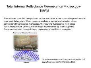
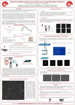
T1 Absorption Fluorescence Fast relaxation Intersystem crossing S0-S1 mixing Radiative transition [1] Microscope Objective 60x,NA=1.4, oil CCD image plane Dichroic mirror Total Internal Reflection (TIR) Filter CCD camera 532 nm Nd:YAG Laser Q=10° Q=0° Q=20° Q=30° 1.5X magnification element Total Internal Reflection Fluorescence Microscopy of Single Rhodamine B Molecules Mustafa Yorulmaz(1), Alper Kiraz(1), A.Levent Demirel(2) (1)Department of Physics, Koç University, Rumelifeneri Yolu, 34450 Sariyer, Istanbul, Turkey (2)Department of Chemistry, Koç University, Rumelifeneri Yolu, 34450 Sariyer, Istanbul, Turkey Motivation Photophysical Properties During the past 15 years, single molecule studies grew rapidly, particularly in its application to biological systems and chemical processes. Single molecules have been used as markers and local probes for local nano physical and chemical properties of molecular processes in their environments. By using the techniques of high resolution fluorescence microscopy, it is possible to track the location of single molecules in amorphous hosts. In crystalline environments, the fluorescence images of single molecules reveal the three dimensional dipole orientations. With the help of annular illumination, these experiments allow imaging single molecules having dipole moments in a direction perpendicular to the substrate as well as parallel. Moving as well as stationary molecules can be observed by Total Internal Reflection Fluorescence (TIRF) microscopy. The diffusion properties of molecules in different polymeric hosts can be understood by observing the dynamics of single molecules. Here we analyzed single Rhodamine B molecules embedded in polymer thin films. We observed different patterns of emission which provides information about the three dimensional dipole moment orientation of molecules. They are doughnut-like structures, rings, asymmetric rings, or spots. We also imaged the diffusion of single Rhodamine B molecules. Blinking • The molecule undergoes an intersystem crossing to its lowest triplet state T1. The transition accompanies by a spin flip of the excited electron and is thus symmetrically disfavored. • Intersystem crossing rates are low, one crossing for every 105-106 excitations. • However the average lifetime of triplet state is much higher then the fluorescence lifetime. The average fluorescence lifetime of Rhodamine B is ~2 ns[2]. Photobleaching • An irreversible chemical reaction that occurs while the electronis in its excited state. It results with final disappearanceof moleculefrom observation. Typical fluorescentdye molecules survive about 105to106 excitation cycles until photodestruction,although thisnumber can vary widely andstrongly depends on the natureof the embedding medium [3]. Blinking and Photobleaching Rhodamine B, PMMA S1 Blinking 2 3 1 Photobleaching S0 6 5 4 There is a 3 seconds time interval between consecutive images. Fluorescence Emission Spectrum of Rhodamine B PMMA Polymer Thin Films Image of a SingleDipole Depending on the Orientation PMMA (C5O2H2)nis a clear, colorless polymer of methyl methacrylate. PMMA (Poly (methyl methacrylate)) thin films areamorphous hosts in which Rhodamine B molecules don not have a preferred orientation. For an oscillating electric dipole with amplitude vector , in a medium with refractive index n0 and distance z0 above a planar interface, the electric field amplitude of the dipole at position z > 0 is given by the plane-wave representation [1]; Sample Preparation • 3mg/ml PMMA in chloroform (CHCl3) solution is prepared • A small amount of 0.2 nM Rhodamine B in methanol was added to the solutions. • Glass substrates were put into the UV-Ozone Cleaner for 30 minutes before spin coating. • Solutions were spin-coated onto glass substrates at 2000 rpm, for 1 minute. • The solvents were dried in a pressure oven. Fourier Transform Experimental Setup Images obtained with electromagnetic calculations and characterization of optical system. A continuous wave laser (l=532nm) was used for excitation in inverted geometry. A high numerical aperture microscope objective (N.A.=1.4, 60x oil) was used for excitation. The collimated laser beam was focused to the back aperture of the microscope objective for wide-field illumination. The angle of incidence of the laser beam to the polymer-air interface was further adjusted to observe total internal reflection. The achieved “annular illumination” enabled the excitation of molecules with dipole orientations perpendicular to the substrate as well as parallel [4]. The emitted fluorescence was collected by the same microscope objective (epi-fluorescence set up) and transmitted through a dichroic mirror, a 1.5x magnification element and a bandpass filter. TIRF microscopy images were detected by a thermoelectrically cooled charge coupled device (CCD) camera. Diffusion Properties of Single Molecules Diffusion Properties of Single Molecules Consecutive TIRF microscopy images reveal the specific path followed by a single Rhodamine B molecule diffusing in PMMA film. Consecutive TIRF microscopy images reveal the specific path followed by a single Rhodamine B molecule diffusing in PMMA film. There is a 0.2 second time interval between consecutive images. There is a 0.2 second time interval between consecutive images. Single Molecules in Polymer Host PMMA We observed diffusing as well as stationary single molecules. Stationary single molecules revealed different emission patterns (doughnut-like structures, rings, asymmetric rings, or spots) due to the different dipole orientations (Fig.1). While some shapes possessed circular symmetry, some shapes were circularly asymmetric. The asymmetry was due to the tilt of the emission dipole with respect to the optical axis. The observed variety of images was explained by calculating the emission pattern of a dipole located below a dielectric-air interface [1]. 3 2 1 Trajectories fallowed by a diffusing single molecule Conclusions Using total internal reflection fluorescence microscopy, we determined the 3-D dipole orientation and diffusion properties of single Rhodamine B molecules embeded in a PMMA thin film. We are planning to use this method to explore the morphology of different polymeric thin films. References: [1] M.A. Lieb, Single Molecule Orientations Determined by Direct Emission Pattern Imaging, J. Opt. Soc. Am. B./Vol. 21, No:6/June 2004 [2] M. Böhmer, J. Enderlein, Orientation Imaging of Single Molecules by Wide-field Epifluorescence Microscopy, J. Opt. Soc. Am. B./Vol. 20, No:3 /March 2003 [3] Ch. Zander, J. Enderlein, R. A. Keller, Single Molecule Detection in Solution, WILEY-VCH, 2002. [4] R. J. P. Zimmermann, C. Hettich, I. Gerhardt, A. Renn, V. Sandoghdar, “Aligned Terrylene Molecules in a Spin Coated Crystalline Film of p-Terphenyl”, Chemical Physics Letters, January 2004. Acknowledgements: This work was supported by the Scientific and Technological Research Council of Turkey (Grant No. TÜBİTAK-107T211). A. Kiraz acknowledges the financial support of the Turkish Academy of Sciences in the framework of the Young Scientist Award program (Grant No. A.K/TÜBA-GEBİP/2006-19). Single molecule images obtained by TIRF microscopy Koç UniversityNano-Optics Research Laboratory, Rumeli Feneri Yolu, Sariyer, Istanbul 34450 Turkey • myorulmaz@ku.edu.tr