Download
1 / 1
10 likes | 113 Views
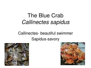
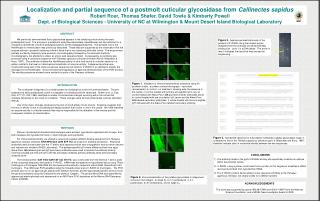
Localization and partial sequence of a postmolt cuticular glycosidase from Callinectes sapidus Robert Roer, Thomas Shafer, David Towle & Kimberly Powell Dept. of Biological Sciences - University of NC at Wilmington & Mount Desert Island Biological Laboratory. NSF. ABSTRACT

E N D
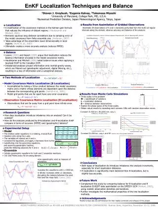
Localization and partial sequence of a postmolt cuticular glycosidase from Callinectes sapidus Robert Roer, Thomas Shafer, David Towle & Kimberly Powell Dept. of Biological Sciences - University of NC at Wilmington & Mount Desert Island Biological Laboratory NSF ABSTRACT We previously demonstrated that a glycosidase appears in the calcifying cuticle during the early postecdysial hours. The enzyme is a putative N-acetylhexosaminidase (HexNAcase) and has affinities for p-nitrophenyl derivatives of both N-acetylglucosamine and N-acetylgalactosamine. The possible role of the HexNAcase in mineralization was previously described. These data are supported by the observation that the enzyme activity in postmolt calcifying cuticle is twice that in non-calcifying arthrodial cuticle. The enzyme was partially purified by Sephacryl size-exclusion chromatography followed by Concanavalin A affinity chromatography, but attempts to obtain an amino acid sequence failed. Consequently, an antibody was produced using a consensus sequence from Penaeus japonicus cuticular chitinase (Pjchi2; Watanabe & Kono, 1997). The antibody inhibited the HexNAcase activity in vitro, and bound to cuticular sections in a pattern similar to that seen in histochemical localization of enzyme activity. A degenerate primer was constructed using part of the same consensus sequence and used for 3'-RACE in an attempt to amplify the message for the HexNAcase. One prominent band appeared on agarose electrophoresis of the PCR product. The resulting sequence showed some similarity to parts of the Penaeus chitinase. Figure 3.Agarose gel electrophoresis of the product of 3’-RACE using the forward primer designed from the chitinase conserved GlcNAc binding site. Lane 1 is a DNA ladder. The arrow in lane 2 indicates the band that was excised for sequencing. Figure 1.Validation of immunohistochemical procedure using 0h-postmolt cuticle. a) phase contrast micrograph – epicuticle (arrowheads); b) control - no treatment, showing auto-fluorescence in the cuticle; c) control treated with primary anti-peptide IgYs only; d) control treated with Alexa-Fluor 488 goat anti-chicken antibodies alone; e) control treated with pre-inoculation IgY serum and the Alexa-Fluor 488-labeled secondary antibodies; f) cuticle treated with the anti-peptide IgYs followed with the Alexa-Fluor-labeled secondary antibody. INTRODUCTION The crustacean integument is a model system for studying the control of biomineralization. The pre-ecdysial and early postecdysial cuticle is incapable of nucleating calcium carbonate. Shafer et al. (J. Exp. Zool. 271:171-182, 1995) identified a number of biochemical changes occurring within the cuticle that correlate temporally to the onset of nucleation. These changes were termed postecdysial cuticular alteration (PECA). One of the major changes observed is the loss of ConA affinity of two mucins. Evidence suggests that this loss of affinity is due to a postecdysial deglycosylation that occurs in situ in the cuticle. We have identified an enzyme activity in cuticular extracts that may be responsible for the alteration of the mucins and the consequent initiation of mineralization. METHODS Pieces of postecdysial dorsobranchial carapace were excised, hypodermis separated with forceps, and both carapace and hypodermis frozen in liquid nitrogen and lyophilized. For immunohistochemisty, we utilized a conserved, putative GlcNAc-binding sequence from Penaeus japonicus cuticular chitinase(KRH NFD GLD LDW EYP VC) as a basis for antibody production.Polyclonal antibodies were constructed from the 17 amino acid sequence which was conjugated to bovine serum albumin and injected into chickens (AVES Laboratory). The epitope-specific IgY’s were affinity-purified from eggs. Alexa-Fluor 488-labeled goat anti-IgY secondary antibodies were used to localize the antibody binding. Controls included pre-immune IgY’s with the secondary antibody, primary antibody alone and secondary antibody alone. The forward primer, GAY TGG GAR TAY CCI GTI TG, was constructed from the terminal 7 amino acids of the conserved sequence and used for 3’-RACE. mRNA was extracted from hypodermal tissue using TRIzol (Invitrogen) or Promega’s Total RNA Kit, and reverse-transcribed to make first-stand cDNA (SuperScript II kit, Invitrogen). The cDNA was PCR-amplified using the forward primer and a 3’-RACE kit (Invitrogen). The PCR product was run on an agarose gel, stained with ethidium bromide, and the appropriate band(s) were cut from the gel and extracted using the QiaQuick kit and protocol (Qiagen). The gel-purified cDNA was quantified by agarose gel electrophoresis and sequenced on an ABI Prism 3100 sequencer at the Marine DNA Sequence Center at MDIBL. Figure 3.Nucleotide sequence of the putative Callinectes sapidus glycosidase (csgly-1) compared to that of the Penaeus japonicus chitinase (pjchi-2; Watanabe and Kono, 1997). Asterisks indicate sites of nucleotide identity between the two sequences. • CONCLUSIONS • 1) The antibody made to the pjchi-2 GlcNAc binding site specifically localizes an epitope within the postmolt cuticle. • 2) 3’-RACE, using a primer constructed from a portion of this sequence, amplified a cDNA reverse-transcribed from hypodermal mRNA. • The 3’-RACE product demonstrates some sequence similarity to the Penaeus japonicus chitinase, but clearly codes for a different protein. Figure 2. Immunolocalization of the putative glycosidase in integument at various molt stages: a) stage D3, b) 0 h postecdysis, c) 3 h postecdysis, d) 8 h postecdysis, and e) stage C4. ACKNOWLEDGEMENTS This work was supported by grants IBN 98-07804 and IBN 0114597 from the National Science Foundation, and a MDIBL New Investigator Award to RDR