Download
1 / 33
340 likes | 729 Views
Gastrulation, Neurulation and Folding. Dr Rania Gabr. OBJECTIVES. By the end of this lecture , the student should be able to: Define Gastrulation Describe the formation of the primitive streak Describe the formation of the intraembryonic mesoderm and the Trilaminar disc

E N D
Gastrulation, Neurulation and Folding Dr Rania Gabr
OBJECTIVES By the end of this lecture , the student should be able to: • Define Gastrulation • Describe the formation of the primitive streak • Describe the formation of the intraembryonic mesoderm and the Trilaminar disc • Explain the formation , function and fate of the notochord • Define Neurulation • Describe the formation of the neural plate, groove, fold , crest and canal • Understand the process of folding its timing and results
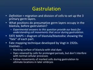
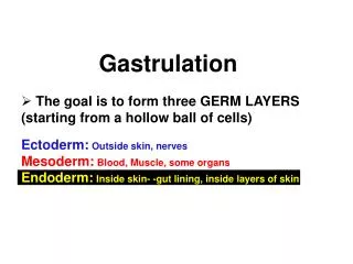
The Third Week The significant event of third week is Gastrulation Gastrulation: is the process of formation of the 3 germ layers (ectoderm, mesoderm & endoderm).
Gastrulation The process by which the bilaminar disc is converted into a trilaminar disc It is the beginning of morphogenesis (formation of body form) Consists of formation of the primitive streak, the three germ layers & the notochord Embryo is referred to as a Gastrula
Primitive Streak The primitive streak results from proliferation of the epiblastic cells in the median plane, in the caudal halfof the epiblast, and lies along the cranio-caudal axis. Its cranial end forms the primitive node A groove, primitive groove, appears in the primitive streak, which continues with a small depression, primitive pit, in the primitive node.
A circular thickening appears in the hypoblastnear the cranial end, in the midline, to form the prechordalplate ( oropharyngeal membrane), that marks the future site of mouth A circular thickening appears in the hypoblast caudal to primitive streak in the midline to form the cloacal membrane, the future site of the anus
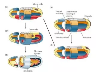
Formation of Intraembryonic Mesoderm The epiblastic cells from the primitive streak (groove) proliferate to form mesenchymaltissue The newly formed cells invaginateand migrateventrally, laterally & cranially between the epiblast and hypoblast & organize to form the intraembryonic mesoderm
Formation of Intraembryonic Mesoderm cont’d Intraembryonic mesoderm merges with the extra-embryonic mesoderm at the periphery of the embryonic disc By the end of3rd week, mesoderm lies between embryonic ectoderm and endoderm everywhereEXCEPT in the region ofprechordal plate andcloacal membrane, as the embryonic ectoderm & endoderm are fused at these regions
Formation of Intraembryonic Mesoderm cont’d Some mesenchymal cells displace the hypoblasts forming the embryonic endoderm Cells remaining in the epiblast form the embryonic ectoderm
Each of the three germ layers gives rise to specific tissues and organs Thus the EPIBLASTgives rise to all three germ layers,Ectoderm, Mesoderm, Endodermin the embryo
Fate of Primitive Streak Actively forms mesoderm until the early part of 4th week Then it starts regressing and becomes an insignificant structure in the sacrocooccygeal regions Normally it degenerates and disappears by the end of 4th week Remnants may persist and give rise to a large tumor called SacrococcygealTeratomas
Notochord A rod of mesenchymal cells located cranially, in the midline, extending between the primitive node and the prechordal plate
Formation of Notochord Mesenchymal cells migrate cranially from the primitive pit towards the prechordal plate, and form a rod like notochordal process The notochordal process becomes canalized forming a hollow tube, the notochordal canal, communicating with the primitive pit.
Formation of Notochord cont’d The floor of the tube and the underlying endoderm break down, forming a notochordal plate The notochordal plate becomes continuous with the endodermal layer.
Formation of Notochord cont’d A temporary communication is established between the amniotic cavity and the yolk sac, termed the neurenteric canal.
Functions of Notochord Defines primordial axis of the embryo Provides rigidity to the embryo Serves as a basis for the development of the axial skeleton Indicates the future site of the vertebral bodies/column Regulates differentiation of surrounding structures including the overlying ectoderm (neural plate) and mesoderm (somites).
Fate of Notochord Degenerates and disappears as the bodies of the vertebrae develop, but it persists as the nucleus pulposusof each intervertebral disc Remnants of notochordal tissue give rise to tumors called Chordomas
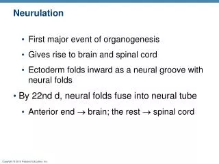
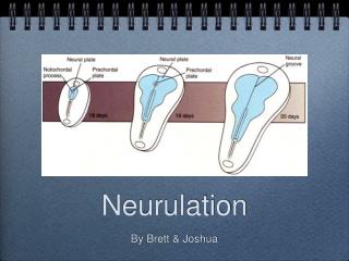
Ectodermal DerivativesThe Neurulation • It is the process by which the neural tube is formed. • The stages of neurulationinclude the formation of: • Neural plate • Neural groove • Neural folds& their fusion • Neural crest cells • Neural tube • Begins during early part of the 4th week (22-23 days) • Ends by the end of 4th week (27 days) • Is induced by the notochord
The Neurulation • Under the inducing effect of the developing notochord, the overlying ectodermal cells thickens to form the neural plate
The neural plate first appears: Cranial to the primitive node and Dorsal to the developing notochord & the mesodermadjacent to it
As the notochord forms & elongates: The embryonic disc elongates and becomes club-shaped The neural plate broadens and extendscraniallyas far as the buccopharyngeal membrane, and later on grows beyond it
On 18th day: the neural plate invaginates to form neural groove & neural folds Neural fold
Some neuroectodermal cells along the crest of the neural fold differentiate as theneural crest cells. Neural crest cells Neural fold
By the end of 3rd week, the neural folds move to the midline and fuse to form the neural tube The fusion begins in the future cervical region and then extends both in cranial and caudal direction
The neural tube separates from the surface ectoderm, lies in the midline, dorsal to the notochord
Neural tube is open at both ends, communicating freely with the amniotic cavity. The cranial opening, the rostral neuroporecloses at about 25th day & the caudal neuroporecloses at about the 27th day
The cranial ⅓ of the neural tube represent the future brain The caudal ⅔ represents the future spinal cord
Folding Of Embryo • Folding means conversion of the flat trilaminar embryonic disc into a cylindrical embryo. Time: Folding of the embryo begins by the end of the 3rd week. It is completed by the 4th week.
Folding of the embryo is due to rapid growth of the embryo specially the nervous system. The head folds first then the tail . At the same time, side to side folding occurs.