Download
1 / 44
440 likes | 580 Views
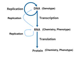
From DNA to Protein: Genotype to Phenotype. One Gene, One Polypeptide. A gene is defined as a DNA sequence that encodes information. In the 1940s, Beadle and Tatum showed that when an altered gene resulted in an altered phenotype, that altered phenotype always showed up as an altered enzyme.

E N D
One Gene, One Polypeptide • A gene is defined as a DNA sequence that encodes information. • In the 1940s, Beadle and Tatum showed that when an altered gene resulted in an altered phenotype, that altered phenotype always showed up as an altered enzyme. • Their results suggested that mutations cause a defect in only one enzyme in a metabolic pathway. • This lead to the one-gene, one-enzyme hypothesis.
One Gene, One Polypeptide • The gene–enzyme connection has undergone several modifications. Some enzymes are composed of different subunits coded for by separate genes. • This suggests, instead of the one-gene, one enzyme hypothesis, a one-gene, one-polypeptide relationship. • Today, we know some genes encode functional RNA molecules, such as ribozymes.
DNA, RNA, and the Flow of Information • The expression of a gene takes place in two steps: • Transcription makes a single-stranded RNA copy of a segment of the DNA. • For functional RNAs, this is the final step. • Translation uses information encoded in the RNA to make a polypeptide.
DNA, RNA, and the Flow of Information • RNA (ribonucleic acid) differs from DNA in three ways: • Single stranded. • The sugar in RNA is ribose, not deoxyribose. • RNA has uracil instead of thymine. • RNA can base-pair with single-stranded DNA (adenine pairs with uracil instead of thymine) and also can fold over and base-pair with itself.
DNA, RNA, and the Flow of Information • Messenger RNA, or mRNA moves from the nucleus of eukaryotic cells into the cytoplasm, where it serves as a template for protein synthesis. • Transfer RNA, or tRNA, is the link between the code of the mRNA and the amino acids of the polypeptide, specifying the correct amino acid sequence in a protein.
Transcription: DNA-Directed RNA Synthesis • In normal prokaryotic and eukaryotic cells, transcription requires the following: • A DNA template for complementary base pairing • The appropriate ribonucleoside triphosphates (rATP, rGTP, rCTP, and rUTP) to act as substrates • The enzyme RNA polymerase
Transcription: DNA-Directed RNA Synthesis • The first step of transcription, initiation, begins at a promoter, a special sequence of DNA. • There is at least one promoter for each gene to be transcribed. • The RNA polymerase (synthesizes RNA during transcription) binds to the promoter region when that protein is needed by the cell.
Transcription: DNA-Directed RNA Synthesis • After binding, RNA polymerase unwinds the DNA and reads the template in the 3¢-to-5¢ direction (elongation). • The new RNA elongates from its 5¢ end to its 3¢ end; thus the RNA transcript is antiparallel to the DNA template strand. • RNA polymerization is always 5’ to 3’ (needs free 3’ –OH to add nucleotide).
Figure 12.4 (Part 3) DNA is Transcribed in RNA Particular base sequences in the DNA specify termination – the signal that the end of the gene has been reached and transcription can terminate.
The Genetic Code • A genetic code relates genes (DNA) to mRNA and mRNA to the amino acids of proteins. • mRNA is read in three-base segments called codons. • The 64 possible codons code for only 20 amino acids and the start and stop signals. • Each codon is assigned only one amino acid.
Preparation for Translation: Linking RNAs, Amino Acids, and Ribosomes • The codon in mRNA and the amino acid in a protein are related by way of an adapter—a specific tRNA molecule. • tRNA has three functions: • It carries an amino acid. • It associates with mRNA molecules. • It interacts with ribosomes.
Preparation for Translation: Linking RNAs, Amino Acids, and Ribosomes • A tRNA molecule has 75 to 80 nucleotides and a three-dimensional shape. • The shape is maintained by complementary base pairing and hydrogen bonding. • The three-dimensional shape of the tRNAs allows them to combine with the binding sites of the ribosome.
Preparation for Translation: Linking RNAs, Amino Acids, and Ribosomes • At the 3¢ end of every tRNA molecule is a site to which its specific amino acid binds covalently. • Midpoint in the sequence are three bases called the anticodon. • The anticodon is the contact point between the tRNA and the mRNA. • The anticodon is complementary (and antiparallel) to the mRNA codon. • The codon and anticodon unite by complementary base pairing.
Figure 12.7 Transfer RNA Anticodon
Preparation for Translation: Linking RNAs, Amino Acids, and Ribosomes • The ribosome is a complex protein assembly where protein synthesis takes place. • The ribosome binds to the mRNA, and then the correct transfer RNA comes in and binds to bring in the correct amino acid – thus building the protein chain. • Each ribosome has two subunits: a large and a small one.
Preparation for Translation: Linking RNAs, Amino Acids, and Ribosomes • The ribosome validates the three-base-pair match between the mRNA and the tRNA. • If hydrogen bonds have not formed between all three base pairs, the tRNA is ejected from the ribosome.
Figure 12.10 The Initiation of Translation The ribosome attaches to the mRNA at a special ribosome recognition sequence upstream of the start sequence AUG. The start codon (AUG) designates the first amino acid in all proteins = methionine.
Figure 12.11 Translation: The Elongation Stage The ribosome helps form a peptide bond between the last amino acid of the growing protein and the amino acid attached to the incoming tRNA.
Figure 12.12 The Termination of Translation When a stop codon—UAA, UAG, or UGA—enters the ribosome, it signals the ribosome to release the formed protein.
Posttranslational Events • Two posttranslational events can occur after the polypeptide has been synthesized: • The polypeptide may be moved to another location in the cell, or secreted. • The polypeptide may be modified by the addition of chemical groups, folding, or trimming.
Figure 12.14 Destinations for Newly Translated Polypeptides in a Eukaryotic Cell
Posttranslational Events • As the polypeptide chain forms, it folds into its 3-D shape. • The amino acid sequence also contains an “address label” indicating where in the cell the polypeptide belongs.
Posttranslational Events • Most proteins are modified after translation. • These modifications are often essential to the functioning of the protein. • Three types of modifications: • Proteolysis (cleaving) • Glycosylation (adding sugars) • Phosphorylation (adding phosphate groups)
Mutations: Heritable Changes in Genes • Mutations are heritable changes in DNA—changes that are passed on to daughter cells. • Multicellular organisms have two types of mutations: • Somatic mutations are passed on during mitosis, but not to subsequent generations. • Germ-line mutations are mutations that occur in cells that give rise to gametes.
Mutations: Heritable Changes in Genes • Point mutations result from the addition or subtraction of a base or the substitution of one base for another. • Point mutations can occur as a result of mistakes during DNA replication or can be caused by environmental mutagens. • Because of degeneracy (redundancy) in the genetic code, some point mutations result in no change in the amino acids in the protein.
Mutations: Heritable Changes in Genes • Some mutations cause an amino acid substitution. • An example in humans is sickle-cell anemia, a defect in the b-globin subunits of hemoglobin. • The b-globin in sickle-cell differs from the normal by only one amino acid. • These mutations may reduce the functioning of a protein or disable it completely.
Mutations: Heritable Changes in Genes • Some mutations are base substitutions that substitute a stop codon. • The shortened proteins are usually not functional.
Mutations: Heritable Changes in Genes • A frame-shift mutation consists of the insertion or deletion of a single base in a gene. • This type of mutation shifts the code, changing many of the codons to different codons. • These shifts almost always lead to the production of nonfunctional proteins.
Mutations: Heritable Changes in Genes • Spontaneous mutations are permanent changes, caused by any of several mechanisms. • Induced mutations are changes caused by some outside agent (mutagen).