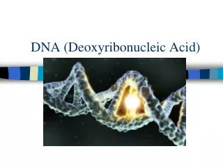
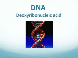
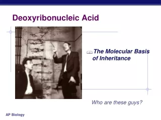
Deoxyribonucleic Acid
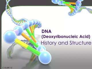
Deoxyribonucleic Acid. (DNA). The double helix. Nitrogenous Bases and Pentose Sugars. Purine and Pyrimidine Structure. (1) Pyrimidines are planar (2) Purines are nearly planar (3) Numbering is different . Numbering Is Different. Bases Have Tautomeric Forms. Uracil. Glycosidic bond.

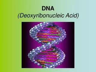
Deoxyribonucleic Acid
E N D
Presentation Transcript
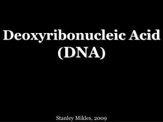
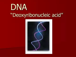
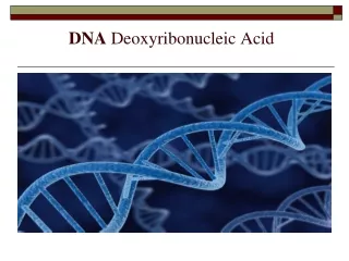
Deoxyribonucleic Acid (DNA)
Purine and Pyrimidine Structure (1) Pyrimidines are planar (2) Purines are nearly planar (3) Numbering is different
Bases Have Tautomeric Forms Uracil
Glycosidic bond Nucleosides vs. Nucleotides
Ester bonds Dinucleotides and Polynucleotides
G=C Watson-Crick Base Pairs A=T
Other Base Pairs Are Possible Watson-Crick, Reverse Watson-Crick, Hoogsteen, Reverse Hoogsteen, Wobble, Reverse Wobble Homo Purines Hetero Purines
Base Pairing Can Result in Alternative DNA Structures Triplex Tetraplex Cruciform Hairpin Loop
Periodicity: A pair of strong vertical arcs (C & N atoms) indicate a very regular periodicity of 3.4 Å along the axis of the DNA fiber. • Astbury suggested that bases were stacked on top of each other "like a pile of pennies". • Helical nature: Cross pattern of electron density indicates DNA helix and angles show how tightly it is wound. • Diameter: lateral scattering from electron dense P & O atoms.
DNase can only cleave external bond demonstrating periodicity
Hydrophilic Hydrophobic Complementarity Watson and Crick Model (1953) • 2 long polynucleotide chains coiled around a central axis • Bases are 3.4 Å (0.34 nm) apart on inside of helix • Bases flat & lie perpendicular to the axis • Complete turn = 34 Å • 10 bases/turn • Diameter = 20 Å • Alternating major and minor grooves
Base Pairing Results from H-Bonds Only A=T and GC yield 20 Å Diameter
Biologically Significant Form = B-DNA Low Salt = Hydrated, 10.5 bp/turn
Side-view Top-view A- DNA Exists Under High Salt Conditions Base pairs tilted, 23 Å, 11bp/turn
Z-DNA Is a Left-Handed Helix Zig-zag conformation, 18 Å, 12 bp/turn, no major groove
Denaturation of DNA Strands and the Hyperchromic Shift • Denaturation (melting) is the breaking of H, but not covalent, bonds in DNA double helix duplex unwinds strands separate • Viscosity decreases and bouyant density increases • Hyperchromic shift – uv absorption increases with denaturation of duplex • Basis for melting curves because G-C pairs have three H bonds but A-T pairs have only two H bonds • Duplexes with high G-C content have a higher melting temperature because G-C pairs require a higher temperature for denaturation
Molecular Hybridization • Reassociation of denatured strands • Occurs because of complementary base pairing • Can form RNA-DNA Hybrids • Can detect sequence homology between species • Basis for in situ hybridization, Southern and Northern blotting, and PCR
Reassociation Kinetics • Derive information about the complexity of a genome • To study reassociation, genome must first be fragmented (e.g. by shear forces) • Next, DNA is heat-denatured • Finally, temperature is slowly lowered and rate of strand reassociation (hybridization) is monitored
Initially there is a mixture of unique DNA sequence fragments so hybridization occurs slowly. As this pool shrinks, hybridization occurs more quickly • C0t1/2= half-reaction time or the point where one half of the DNA is present as ds fragments and half is present as ss fragments • If all pairs of ssDNA hybrids contain unique sequences and all are about the same size, C0t1/2is directly proportional to the complexity of the DNA • Complexity = X represents the length in nucleotide pairs of all unique DNA fragments laid end to end • Assuming that the DNA represents the entire genome and all sequences are different from each other, then X = the size of the haploid genome
Maximum denaturation = 100% single stranded 50% double, 50% single stranded Double stranded The Hyperchromic Shift (Melting Curve Profile) Tm = temperature at which 50% of DNA is denatured
High G-C Content Results in a Genome of Greater Bouyant Density
100% ssDNA 100% dsDNA Ideal C0t Curve