Download
1 / 22
260 likes | 487 Views
Myosin Contracts Skeletal Muscle . Jonathan P. Davis, Ph.D. Assistant Professor Office/Lab Phone 247-2559 Email: davis.812@osu.edu. Department of Physiology and Cell Biology, The Ohio State University, 400 Hamilton Hall. Muscle.

E N D
Myosin Contracts Skeletal Muscle Jonathan P. Davis, Ph.D. Assistant Professor Office/Lab Phone 247-2559 Email: davis.812@osu.edu Department of Physiology and Cell Biology, The Ohio State University, 400 Hamilton Hall
Muscle Skeletal Cardiac Smooth
Thick Filament or Myosin Regulation In these cases: Myosin needs to be “activated” before it can interact with actin or move cargo
Actin Binding Proteins Like Tropomyosin Regulate Myosin Thin Filament Regulation “Blocked State” “Open or M State” “Closed State” Strong Hydrophobic Myosin Binding Sites Weak Electrostatic Myosin Binding Sites Tropomyosin “Rocks and Rolls”
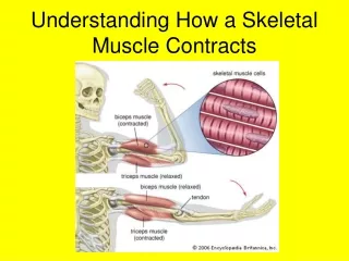
Structure of Skeletal (striated) Muscle Comprised of fibers (cells) Each fiber contains many myofibrils in parallel Each myofibril contains many sarcomeres in series Striations due to characteristic banding pattern of sarcomere Electron micrograph of Skeletal Muscle Fiber
Sarcomere Composed of Overlapping Thin and Thick Filaments The Sarcomere
CROSS-BRIDGES PROJECT FROM THICK TO THIN FILAMENTS Z Line H-Zone Cross-bridge
The Thin Filament Is Composed Primarily of Actin but Also Contains Tropomyosin and the TroponinComplex Complex The Troponin Complex Contains Three Proteins • Troponin C – Binds Calcium • Troponin I – Inhibits Cross-Bridge Binding • Troponin T – Binds Tropomyosin
T-tubules and the Sarcoplasmic Reticulum The transverse tubules bring action potentials into the interior of the skeletal muscle fibers, so that the wave of depolarization passes close to the sarcoplasmic reticulum, stimulating the release of calcium ions. The extensive meshwork of sarcoplasmic reticulum assures that when it releases calcium ions they can readily diffuse to all of the troponin sites.
ACTION POTENTIAL CAUSES RELEASE OF CALCIUM FROM SR Cell Membrane Ryanodine Receptor Sarcoplasmic Reticulum T -Tubule SR Ca2+ ATPase Dihydropyridine Receptor Calsequestrin Calcium
Mechanism of Skeletal Muscle Activation by Ca2+ **Tm can occupy 2 positions: “off” and “on” state 1) “off” state- absence of Ca2+ Tm blocks myosin binding 2) “on” state- Ca2+ binds to TnC Tm moves toward center of actin Myosin binding sites exposed Muscle contracts
Regulation of Striated Muscle Contraction 1) Action Potential 2) Calcium Transient Plasma Membrane Plasma Membrane Sarcoplasmic Reticulum [Ca2+] T-Tubule Sarcoplasmic Reticulum **SR Ca2+ ATPase** Time Calcium 3) Calcium Binds Troponin C 4) Myosin Power Stroke 5) Force Production Actin Actin –Ca2+ Relaxed Tropomyosin Troponin Complex - Ca2+ Actin Myosin Myosin + Ca2+ Myosin Binding Site +Ca2+ Contracted *** ATP Driven Power Stroke & Detachment***
ISOTONIC AND ISOMETRIC CONTRACTIONS Greater the force against which shortening occurs, the slower the velocity of shortening A – 100% Maximal Force B – 75% Maximal Force Force Maximal Velocity (VMAX) C – 50% Maximal Force D – 25% Maximal Force D C = L/T D B C A Muscle Shortening B A Time Muscles exhibit > 200-fold variation in maximum velocity of shortening. Why? Maximum velocity of shortening Reflects speed of cross-bridge cycling Is actomyosin ATPase activity Is determined by differences in the myosin molecule
RELATIONSHIP OF ELECTRICAL TO MECHANICAL EVENT IN SKELETAL MUSCLE CONTRACTION Calcium Transient (Shortening or Force Generation)
FORCE DEVELOPMENT IN AN ISOMETRIC CONTRACTION AS A FUNCTION OF STIMULUS FREQUENCY
ACTIVE, PASSIVE AND TOTAL FORCE VERSUS MUSCLE LENGTH Isometric contraction at each length In the body Skeletal muscle operates at plateau of length-force relation Cardiac muscle operates on the ascending limb of length-force relation
CLASSIFICATION OF SKELETAL MUSCLE FIBERS Classification system of muscle fibers is based on: Rate of ATP utilization and capacity to re-synthesize ATP Physiological implications of these parameters Muscles are heterogeneous with different proportions of fiber types depending on function
RELATIONSHIP OF MOTOR UNITS TO INNERVATED MUSCLE FIBERS AND RECRUITMENT Fast- glycolytic Fast-oxidative Slow-oxidative
LEVER RELATIONSHIP OF MUSCLE TO BONE AFFECTS FORCE DEVELOPMENT AND VELOCITY
EFFECTS OF FATIGUE ON SKELETAL MUSCLE FIBERS TYPES • What Could be Happening? • Conduction Failure • Energy Metabolism Biproducts • A) Lactic Acid • B) Phosphate and ADP
Geometry of Muscle a. Direction fibers run in the muscle b. Lever system