Download
1 / 14
160 likes | 527 Views
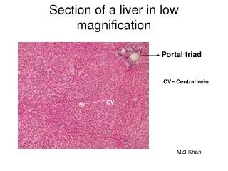
Portal triad. Section of a liver in low magnification. CV= Central vein. CV. MZI Khan. Section of a liver. MZI Khan. Portal Triad. This section of a liver is showing many hepatic lobules centered around central vein. Interlobular connective tissue is not present here,

E N D
Portal triad Section of a liver in low magnification CV= Central vein CV MZI Khan
Section of a liver MZI Khan Portal Triad • This section of a liver is showing many hepatic lobules centered around central vein. • Interlobular connective tissue is not present here, therefore, demarcation of lobules from one another is difficult here.
Section of a Pig Liver MZI Khan • Section of this liver is • Showing: • Benzene-shaped hexagonal hepatic lobules, which are demarcated by connective tissue. • Central vein at the center of lobule. • Portal areas at the angle of lobules.
MZI Khan • Section showing central vein, sinusoids, and hepatic cords. • Hepatic cords consisting of six sided liver cells (hepatocytes). • The hepatocytes seems to be radiating from the central vein towards periphery of the hepatic lobule. • Sinusoids are the open channel of blood in between the hepatocytes. Section of Liver
Section of a liver showing a lobule and surfaces of the hepatocytes MZI Khan • Surfaces of hepatocytes: • (1)Microvillus surfaces that face the • perisinusoidal space, • (2) Surface that border the bile • canaliculi, and • (3) Contact surface between adjacent • hepatocytes where apposed surface • (lateral face). • The hepatocytes have centrally located spherical nucleus with one or more prominent nucleoli. Occasionally binucleated cells may see
Portal areas/canal/triads of liver MZI Khan • Portal triads have: • Hepatic artery • Portal vein, and • Bile duct
Classification of hepatic lobules MZI Khan Classical hepatic lobule Portal lobule Liver acinus
Diagrammatic representation of Classical liver lobule MZI Khan Composition of a lobule already discussed In the previous slides.
The Portal Lobule of liver Portal lobule is a functional unit centered around the bile ductule in the portal area. It is defined as a triangular area consisting of the parenchyma of three adjacent hepatic lobules that are drained by the bile ductule in the portal canal.
Liver acinus Liver acinus is a unit of parenchyma defined in relation to its vascular supply. The liver acinus is a roughly diamond shaped area made of parts of two hepatic lobules supplied by terminal branches of the interlobular portal vein and hepatic artery. The blood vessels course at right angles from the portal area between two hepatic lobules to form backbone of the acinus, and the two central veins are at the two opposing points of the diamond. The liver acinus is divided into three Zones: zone1, 2, and 3.
Diagrammatic representation showing lining cell of the sinusoids and space of Disse MZI Khan • Lining cells of the sinusoids are: • Porous endothelial cell • Von Kupffer cell: Macrophage, & • Perisinusoidal stellate cells or • adipocyte, or Ito cell, which store • vitamin A. • Space of Disse is the space in between endothelium of the sinusoids and hepato -cytes. Here the exchange of the metabolites takes place.
Basic structure of Liver Lobule showing Flow of Blood and Bile MZI Khan
Basic Structure of Liver Lobule Showing Flow of Blood and Bile Flow of blood and bile in the hepatic Lobule is opposite in direction