Download

1 / 11
0 likes | 18 Views
Type 1 diabetes (T1D) is characterized by autoimmune destruction of insulin-producing u03b2-cells in the pancreas, an organ in the abdomen, produces very little or no insulin caused by a complex interaction of genetic and environmental factors. Insulin is a hormone that helps the body to absorb and use glucose and other nutrients from food, store fat, and build up protein and without insulin, blood glucose (sugar) levels become higher than normal. The genetic factors involved consist of multiple susceptibility genes, at least five of which, HLA, INS, CTLA4, PTPN22 and IL2RA/CD25.

E N D
1 Int. J. Biomol. Biomed. International International Journal of Biomolecules and Biomedicine (IJBB) Journal of Biomolecules and Biomedicine (IJBB) ISSN ISSN: : 2221 2221- -1063 (Print) 1063 (Print)2222 Vol. Vol. 3 3, No. , No. 1 1, p. , p. 1 1- -11 http://www.innspub.net http://www.innspub.net 2222- -503X (Online) 503X (Online) 11, 201 , 2013 3 REVIEW PAPEROPEN ACCESS OPEN ACCESS Type 1 diabetes: basis of causes and away of prevention Md. Shamim Hossain*, Mst. Touhida khatun Department of Biotechnology and Genetic Engineering, Faculty of Applied Science and Technology, Islamic University, Kushtia-7003, Bangladesh Article Published: 27 January 2013 Key words: Type 1 diabetes, autoimmune, gene, prevention. Abstract Type 1 diabetes (T1D) is characterized by autoimmune destruction of insulin-producing β-cells in the pancreas, an organ in the abdomen, produces very little or no insulin caused by a complex interaction of genetic and environmental factors. Insulin is a hormone that helps the body to absorb and use glucose and other nutrients from food, store fat, and build up protein and without insulin, blood glucose (sugar) levels become higher than normal. The genetic factors involved consist of multiple susceptibility genes, at least five of which, HLA, INS, CTLA4, PTPN22 and IL2RA/CD25. The excess mortality associated with the complications of type1 diabetes and the increasing incidence of childhood type 1diabetes emphasize the importance of therapeutic strategiesto prevent this chronic disorder. Although no current "cure" exists, recentgenetic data and preliminary trial results suggest T cells asa target for preventive strategies. Another potentially attainabletarget is induction of tolerance to the ß-cell proteinssuch as insulin that are inappropriately recognized. Other strategiesinvolve ß-cell replacement, but currently there are insufficient donor cells available. But researchers revealed that breastfeeding, taking proper exercise, vaccination against causing enterovirous, vitamin D, regulation of maternal diet and keep away from smoking are the best away prevent from type 1 diabetes. This review aims to provide the link of causing type 1 diabetes and the away of prevention to the type 1 diabetes patient. *Corresponding Author: Md. Shamim Hossain shamim_btge@yahoo.com Hossain and Khatun
2 Int. J. Biomol. Biomed. Introduction Genetic factor involved in T1D Type 1 Diabetes(insulin dependent) develops when the Type 1 diabetes is a polygenic disease that many insulin-producing β-cell in the pancreas have been different genes are involved to its expression (Rubio- destroyed and the body unable to produce insulin Cabezas and Argente 2008). On the basis of locus or which causes the increase of glucose levels in the blood combination of loci, it may be dominant, recessive, or (ASH Fact Sheet. 2012). Glucose is a sugar that the between of them. Though the identifying of a complex body produces primarily from the digestion of disease is challenging but differentstrategies have been carbohydrates and levels are controlled by the used in efforts to identify T1D susceptibilitygenes. hormone insulin. Insulin is made and stored in the Among them the most successful were the early studies pancreas and helps glucose to enter the cells where it is of genefrequencies in individuals with T1D compared used as fuel by the body. Without insulin, the body's with controls (Mehers and Gillespie 2008). cells cannot absorb glucose from the blood and as a result, glucose accumulates in the blood, leaving the Human leukocyte antigen body's cells and tissues starved for energy. Type 1 The highly polymorphic humanleukocyte antigen Diabetes usually appears in children and young adults (HLA) on chromosome 6p21.3 express the functional but can occur at any age (ASH Fact Sheet. 2012). differences in how fragments of protein arepresented Studies ofneonataldiabetes express that most cases of to the immune system. In the early 1970s, several diabetes diagnosed before6 months are due to be groups of investigator found that HLA class I are autoimmune, but those diagnosedafter the age of6 associatedwith T1D (Singal et al., 1973; Cudworth and months have the geneticcharacteristics ofT1D (Edghill Woodrow 1975; Mehers and Gillespie 2008). Later, et al., 2006). Islet autoimmune β-cell destruction lymphocyte-defined HLA-Dantigens, HLA class II DR4 occur for the Islet auto antibodies that the and DR3 were shown to be more closelyassociated combinations ofthis antibodies to insulin (IAA), with T1Dand the combination of two susceptiblealleles (Bottazzo et al., 1985) glutamicacid decarboxylase together DR3/DR4 which produced a higher risk (GADA)and the tyrosine phosphatase IA-2 (Hawkes et genetic combination (Todd et al., 19897). The HLA al., 1996)which found in individuals that at risk or who consists of a lots of genes that are close together and have recently developedT1D.This islet auto antibodies transferredfrom the parent to the child in adjacent are detected at the age of 5years (Bonifacio et al., ‘DNA chunks which are known as linkage 2004) and antibodies to INS (generally the first to disequilibrium. Other naturalhistory ofT1D studies appear) have beendetected as early as 6–12 months have demonstrated that HLA genes areassociated with (Roll et al., 1996).It is thought that diabetes play a key the auto antibodies where IA2-A are associatedwith role for introducing different types of diseases. People specific DR alleles (Williams et al., 2008) IAA are with diabetes are at greater risk of heart disease that associated with DR4 (Achenbach et al., 2004) GADA high glucose levels affect the walls of the arteries are associated with DR3and the recently discovered making them more likely to develop fatty deposits ZnT8 antibodiesare associated with a single base pair which in turn makes it more difficult for the blood to change in the SLC30A8gene Class II HLA has been circulate. It also causes high blood pressure, stroke reported to be associated not only with acute-onset kidney disease, nerve damage leading to limb type 1 diabetes but also with fulminant and slowly amputation, eye damage such as retinopathy and lower progressive type 1 diabetes (Imagawa andHanafusa the levels of the protective HDL cholesterol (ASH Fact 2006; Kobayashi et al ., 1993).It is noted as a report Sheet. 2012). on fulminant type 1 diabetes that high frequencies of class II HLA alleles provide resistance to type 1 diabete Hossain and Khatun
3 Int. J. Biomol. Biomed. (Imagawa et al., 2000). Other studies found that where activation (Bottini et al., 2004). In 2008 Ikegami et al., serological typing of class II HLA showed a higher collectedJapanese and Korean samples for sequencing frequency of the DR4-DQ4 as well as DR2-DQ1 of PTPN22 and revealed the presence of five novel haplotype than in autoimmune type 1 diabetes SNPs in the sample and one of these, the -1123 G>C (Imagawa et al., 2005). SNP in the promoter region, was found to be associated with type 1 diabetes in both Japanese and INS gene Korean populations. In 1983, a second locus of a variable numbertandem repeat of INS promoter region was indentified that is IFIH1 responsible for regulation of INS production and has a The IFIH1 gene encodes helicase C domain 1on link with susceptibility to T1D (Bell et al., 1984). chromosome 2q24.3 which mediates the interferon Alleles of this promoter region are consisted of three response to viral RNA (Von Herrath 2009).The IFIH1 classes and distinguishedby the number ofDNA base polymorphisms associated with type 1 diabetes pair repeats. Class I alleles have570 base pairs, class II (Nejentsev et al., 2009) because of their connection to alleles 1200 base pairs andclass III alleles have 2200 the inflammatory response caused by infectious agents base pairs. Class I alleles have higher sensitivity to such as enteroviruses. This viral infection induce islet T1D, on the other hand protectionfrom T1D is autoimmunity and influence progression to diabetes associated with the class III allele (Bennett et al., (Hyöty and Taylor 2002; Honeyman 2005). In 2008, 1995). INS gene study has been reveal that class I Qu et al.,carried out a study on IFIH1 and genotyped alleles in the pancreas produce higher INS than class five SNPs in 1767 individuals where found association III alleles, but on the contrary class I alleles are with T1D. expressed at 2–3-foldlower levels inthe thymus than class III alleles. For this reason class III allele alter the IL2RA/CD25 selection of T cellsin the thymus and influence Vella et al.in 2005 noted that the interleukin 2 tolerance to INS (Vafiadis et al., 1997). receptoralpha (IL2RA) region on chromosome 10p15 was associated withT1D. IL2RA encodes the alpha- CTLA-4 chain of the IL-2 receptor complex which is CTLA-4 is known as surface molecule which generally responsible for binding IL-2 that is a key player in the activated the T cells that induces a negative signal for T proliferation of regulatory T cells. The IL2RA region cell activation. If the CTLA-4 gene is inheritably has been reported to be linked not only with type 1 changed it expressioncan increase T cell self-reactivity diabetes but also with autoimmune thyroid diseases and thus play an importantrole in causing (Brand et al., 2007) and suggesting that the IL2RA autoimmune diseases such as T1D. In 2003, Ueda et region plays a general role in autoimmunity. Qu et al., al., reveal that sensitivity of this molecule is in 2007 also confirmed that IL2RA has an association responsible for causing T1D. with T1Dand identified two SNPs which increased the risk ofT1D. After that Lowe et al.,analyzed 288 SNPs PTPN22 and identifiedthe SNP ss52580101 to be the most In 2004, Bottini et al. Found that tyrosine phosphatase closely associated with T1D and they found that SNPs associated with susceptibility to T1D, is a lymphoid are poorly associated with T1D when increased soluble protein produced from a genePTPN22 on chromosome IL2RA expression. So it is reveal that T1D-susceptible 1p13.ThePTPN22 gene contributes tosusceptibility to alleles are associated with decreasedconcentrations of T1D by increasing the negative regulationof T cell IL2RA and suggesting a possible biological mechanism Hossain and Khatun
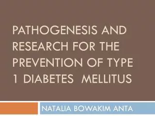
4 Int. J. Biomol. Biomed. for autoimmunity through reduced binding of IL-2 Genome wide association studies which is an essentialcomponent for the survival and The recent advances technology and knowledge in proliferation ofregulatory Tcells suppressors of molecular genetics such as the high density genotyping autoreactivity. chips have made it possible to perform genomewide association (GWA) studies with genotyping of hundreds of thousands of single nucleotide polymorphisms (SNPs) across the genome to denote the genetic basis of common human diseases like as type 1 diabetes. In2007, 2000 individuals and 3000 controls were analyzed for detecting seven major diseases (Todd et al., 2007) where found significant linkages of 12q24, 12q13,16p13, 18p11, 12p13 and 4q27 for T1D. These loci do notoverlap with the previously identified IDDMloci. The further analysiscarriedout of 4000 individuals and 5000controls with T1D where Fig. 1. Peptides of proinsulin from pancreatic b-cells 2997 T1D family trios confirmed 12q24, 12q13, 16p13 are directed by antigen-presenting cells which are then and 18p11(Todd et al., 2007). presented by HLA class II molecules to the T cell receptor on CD4 T cells. Polymorphisms of the CTLA-4 Environmental factor and PTPN22 increase the risk of T cell self-reactivity Though the Identification of environmental factors are and autoreactive CD4 T cells then stimulate cytotoxic very difficult but the most popular candidates are CD8 T cells that attack pancreatic b-cell expressing viruses, with enteroviruses (Hyoty andTaylor 2002), proinuslin peptides through HLA class I molecules on rotavirus (Honeyman et al., 2000) and rubella being their cell surface (Mehers and Gillespie2008) . suspects. The strongest data todate have supported a role for rubella. Infants generally infected with Vitamin D receptor (VDR) congenital rubella syndrome are said to be at increased Vitamin D has important immunomodulatory riskof type 1 diabetes (Ginsberg et al., 1985).Yet properties and theactive form vitamin D3 (1,25 Finland, where vaccination has effectivelyeradicated dihydroxyvitamin D3) has been investigatedto inhibit rubella, still has one of the highest incidencesof type 1 T cell proliferation (Muller and Bendtzen 1992 ).The diabetes (Peltola et al., 2000). There is also some genetic studies of vitamin D related genes suggest that evidence that some enteroviruses(e.g., Coxsackie B it has a correlation of T1D. Though one study viruses) are less prevalent in countrieswith high expressed that there are no association between T1D incidences of type 1 diabetes (e.g., Finland) thanin and the VDR geneafter examining 97 SNPS in up to countries with low incidences but geographically 3763 T1D families from theUK, USA, Finland and similarpopulations (e.g., Russian Karelia) (Viskari et Romania but in 2007 investigated the VDR gene al., 2005). This observation maybe in keeping with the against T1D and found a link with the association of concept of the hygiene hypothesis (Gale 2002; Bach CYP27B1gene on 12q13.1–q13.3 where the C allele of 2005),which support that environmental exposure to rs10877012was significantly associated with increased microbes, otherpathogens and their products early in risk of T1D (Mehers and Gillespie2008). life promotes innate immuneresponses that suppress atopy and perhaps autoimmunity. In Westerncultures, the developing immune system of the infant is no Hossain and Khatun
5 Int. J. Biomol. Biomed. longerexposed to widespread infection which may ((Mathieu and Badenhoop 2005;Ishida et al., 1988). contribute to thecurrent increases in incidence Insulin secretion is impaired by vitamin D deficiency observed in atopic and autoimmunedisease. and restored by 1,25-(OH)2D3 administration (Bourlon et al., 1996). Active of vitamin D is mediated Prevention from type 1 diabetes through its receptor influences gene transcription and Breastfeeding affects pancreatic b-cell function. Vitamin D deficiency According to a Swedish study, if a mother who has a during pregnancy causes the incidence of autoimmune new infant and a family history of type 1 diabetes, diseases, such as type 1 diabetes. Islet cell insulin breastfeeding for a long period of time may be at lower secretion is reduced in vitamin D-deficient animals and risk of developing type 1 diabetes than those who are if high-dose of vitamin D supplementation is given not (Sadauskaite-Kuehne et al., 2004). Breastfeeding early in life it may protects against type 1 diabetes children tend to grow more slowly and steadily while (Mathieu and Badenhoop 2005). formula-fed babies often grow fast because mother's milk contains fewer calories and protein fragments are Vitamin D can be taken up by two sources one is from too small to stimulate the immune system than food (e.g. fatty fishes and their oils) and another can be formula and thus breastfeeding may protect against the achieved through direct ultraviolet B (UV-B)-mediated development of the antibodies associated with type 1 synthesis in the skin. Sunlight has two types of diabetes (Rewers and Gottlieb 2009). The cow's milk ultraviolet radiation. When sunshine in the UV-B hypothesis is being checked of proper functioning spectrum strikes the skin, it converts a substance in the against type 1 diabetes by the Trial to Reduce type 1 skin called 7-dehydrocholesterol into vitamin D3. The diabetes in the Genetically at Risk (Study design of 7-dehydrocholesterol is a very close precursor to TRIGR., 2007) and the Finnish Intervention Trial for cholesterol. UV-B is the short wavelength light that the Prevention of Type I Diabetes (FINDIA) that causes sunburn and does not penetrate the skin deeply. bovine insulin present in cow's milk triggers islet It does not penetrate glass. If one get sunlight behind a autoimmunity. So while mothers are unable to breast window, while driving etc, will not get vitamin D. feed before the 8 months age baby are suggested to Sunscreens also prevent the formation of vitamin D. give either a formula of extensively hydrolyzed protein The another UV-A radiation has a longer wavelength, (Nutramigen) or a formula based on nonhydrolyzed and penetrates through the outer skin deep down to cow's milk (Enfamil) containing a small amount of the elanocytes, the cells that become cancerous in Nutramigen that develop of diabetes by the age of 10 melanoma cases. years (Rewersand Gottlieb 2009). The recent studies also reveal that longer duration of breastfeeding is Beta cells regeneration associated with lower risk of overweight (Harder et al., Yuval Dor, Ph.D professor of Hebrew University 2005) and type 1 diabetes (Sadauskaite-Kuehne et al., Jerusalem revealed in a study that the first time a high 2004) in later life of the child. rate of glucose converts into energy the cells that are involved influenced beta cells regeneration. Coupled Supplementation of Vitamin D with a mechanism that prevents the immune system Recent studies reveal that vitamin D plays an from attacking beta cells in the first place, the long- important role of pathogenesis and prevention of type 1 awaited finding may help the way to a full cure for type diabetes. The β -cell possesses specific receptors for the 1 diabetes (Meier et al., 2005). Benjamin Glaser, M.D. activated hormone 1,25-dihydroxy vitamin D3 and of Hadassah Medical Center, used a genetic system to vitamin D–dependent calcium-binding proteins destroy 80 percent of the insulin-producing cells in Hossain and Khatun
6 Int. J. Biomol. Biomed. adult mice, rendering the mice diabetic. When the type 1 diabetes direct linear with increasing birth researchers compared these mice with control mice, weight. Thus, the way an expectant mother eats can be they found that those mice with diabetes and elevated expected to have an effect on the future health of her blood glucose levels had regenerated a greater number offspring. So the mother usually controls a family's of new beta cells than mice without diabetes and food will also influence the way her children eat. suggesting that glucose is a key player in beta cell regeneration. The researchers also found that an Pancreas transplantation enzyme, glucokinase are involved the first step in Pancreatic transplantation has offered a successful converting glucose for energy that stimulates beta cells therapeuticapproach for many years (Burke et al., to replicate. This study also showed that regeneration 2004).When type 1 diabetes cannot be controlled or is depends on glucokinase levels instead of glucose levels causing serious problems; the patient may want to and researchers may be able to use drugs to trigger decide to a pancreas transplant. For patients with beta cells to regenerate without exposing the body to severe type 1 diabetes, a pancreas transplant probably elevated glucose levels. Another study also found that a offers the greatest chance of a more "normal" lifestyle, single murine adult pancreatic precursor exists that free from insulin shots. Pancreas transplants are safest can differentiateinto cells with the characteristics of in people who do not have heart or blood vessel islet ß cells (Seaberg et al., 2004 ) where shown that disease. Unfortunately, there are not enough donor the pre-existing ß cells, rather than pluripotent stem pancreases to go around because not enough people cells are the main source of newß cells during adult life sign up to be organ donors, and each pancreas must and after pancreatectomyin mice (Dor et al., 2004). meet strict guidelines. When a whole pancreas is not available, a person can receive a portion of a pancreas Maternal diet from a living relative. On the other hand Islet Family dietary traits and lifestyle may play an transplants are intended to treat advanced type important role for causing of type-1 diabetes within 1diabetes by replacing destroyed islets with new ones families. If a pregnant woman eats too much where no surgery is needed. The islets cells from a carbohydrate, this will raise her insulin levels though deceased donor's pancreas are removed and injected insulin itself cannot cross the placenta from mother to into a major blood vessel of the patient's liver. The islet foetus but insulin produces antibodies can.This cells then begin making insulin. With the use of a antibodies in the foetus increase glycogen and fat combinationof daclizumab, sirolimus and tacrolimus deposits resulting in an abnormally large baby which and islets from morethan 1 donor pancreas per may make favorable for the baby to type-1 diabetes. recipient, success rates of 80% at1 year and 20% at 5 Mother diet may play a vital role on the babies diet years have been reported (Ryan et al., 2005). which is the another example of an elimination of diet trial (Schmid et al., 2004).This randomized, unmasked Exercise feasibility study is expressing the effect of delaying Exercise has a greater beneficial result than dietary exposure to gluten until the age of 1 year (Rewersand modifications or even weight loss on the management Gottlieb 2009). Another study launched among the of blood sugar. Regular taking of exercise reduce the group of Norwegian population by record linkage of risk of obesity and thereby minimize the incidence of the medical birth registry and the National Childhood causing of diabetes types diseases. By losing of weight Diabetes Registry looked at all live births in Norway and increasing physical activity can neutralize the between 1974 and 1998 (1,382,602 individuals) (Stene powerful effect of insulin resistance on progression to et al., 2001) where found that increasing incidence of type 1 diabetes (Fourlanos et al., 2004; Xu et al., Hossain and Khatun
7 Int. J. Biomol. Biomed. 2007). Precise blood glucose control after diabetes Conclusions resulting in β-cell rest and it is believed that it helps to Type 1 diabetes risk is mainly depending upon a preserve residual insulin secretion (Brown and Rother genetic susceptibility, unknown environmental trigger 2008). Before the 19th century, it was known that BG and an uncontrolled autoimmune response that attacks concentrations typically decrease with endurance-type the insulin producing beta cells. Though the scientific exercise in most individuals with diabetes (Michael et community is increasingly ableto define the al., 2006). In someone who already has diabetes, importance of individual genes in susceptibilityto T1D exercise and a nutritionally balanced diet can greatly and diagnose or predict the onset ofdiabetes in an limit the effects of both types 1 and 2 diabetes. Intense individual case but it is now unclear what percentage of exercise draws glucose out of the muscle and after absolute geneticrisk can be measured by combining all then the muscles pulls the glucose out of the the known risk alleles of the genes. The subsequent bloodstream and into the muscle cells. The enzyme lack of insulin of the diabetes patient leads to increased AMP kinase initiates glucose transport from blood to blood and urine glucose. Injection is the most common cells without the use of insulin. This is especially method of administering insulin. Pancreas transplants important and helpful in light of the occurrence of have been used to treat type 1 diabetes; however, this insulin resistance in those at risk for diabetes. Exercise procedure is currently still at the experimental trial is found to increase levels of AMP kinase. During post- stage. Some research has suggested that breastfeeding exercise, carbohydrate intake is necessary to help decreased the risk of type 1 diabetes in later life and replenish liver and muscle glycogen stores and this taking proper exercise and maternal diet may also period, which may last up to 12 to 24 h, insulin contribute to prevent diabetes. Giving children 2000 sensitivity are elevated and there is a high risk of IU of Vitamin D during their first year of life is hypoglycemia in patients with type 1 diabetes .For a associated with reduced risk of type 1 diabetes though patients who tend to manage post-exercise late-onset the actual relationship is unknown. We believe that in hypoglycemia during the night, a complex the near future scientist can know the roles of these carbohydrate (e.g. uncooked corn starch) or a mixed genesand their molecular pathways that are related to snack containing fat and protein may be particularly the risk of T1D and may potentially lead to targeted beneficial at bedtime (Michael et al., 2006). therapies for children for treating or preventing diabetes. Vaccination It has been thought that viruses might play a role in References causing type 1 diabetes by infecting the beta cells of the Achenbach P, Koczwara K, Knopff A, Naserke pancreas. The new research suggests that enteroviral H, Ziegler AG, Bonifacio E. 2004. Mature high- infection of the beta cells in children with type 1 affinity immune responses to (pro) insulin anticipate diabetes may initiate a process whereby the body's the autoimmune cascade that leads to type 1 diabetes. immune system identifies beta cells as 'foreign' and Journal of Clinical Investigation 114, 589–597. rejects them. The beta cells are not destroyed in this disease but enteroviral infection of beta cells reduces Action on smoking and health (ASH) Fact their ability to release insulin. Vaccination in childhood Sheet. 2012. Smoking and diabetes, www.ash.org.uk to prevent enteroviral infections of beta cells might be Bach JF. 2005. Infections and autoimmune diseases. an attractive means to reduce the incidence of both J Autoimmun25, 74-80. common forms of diabetes(Gillespie 2006). Hossain and Khatun
8 Int. J. Biomol. Biomed. Bell GI, Horita S, Karam JH. 1984. A polymorphic Brown RJ, Rother KI. 2008. Effects of beta-cell locus near the human insulin gene is associated with rest on beta-cell function: a review of clinical and insulin-dependent diabetes mellitus. Diabetes 33, preclinical data. Pediatr Diabetes 9, 14–22. 176–183. Burke GW, Ciancio G, Sollinger HW. 2004. Advances in pancreas transplantation. Transplantation Bennett ST, Lucassen AM, Gough SC. 1995. 15, 77, S62-7. Susceptibility to human type 1 diabetes at IDDM2 is determined by tandem repeat variation at the insulin Cudworth AG, Woodrow JC. 1975. Evidence for gene minisatellite locus. Nat Genet 9, 284–292. HL-A-linked genes in ‘juvenile’ diabetes mellitus. Br Med J 3, 133–135. Bonifacio E, Hummel M, Walter M, Schmid S, Ziegler AG. 2004. IDDM1 and multiple family history Dor Y, Brown J, Martinez OI. 2004.Adult of type 1 diabetes combine to identify neonates at high pancreatic beat-cells are formed by self-duplication risk for type 1 diabetes. Diabetes Care 27, 2695–2700. rather than stem-cell differentiation. Nature429, 41- 6. Bottazzo GF, Dean BM, McNally JM, MacKay EH, Swift PG, Gamble DR. 1985. In situ Edghill EL, Dix RJ, Flanagan SE. 2006. HLA characterization of autoimmune phenomena and genotyping supports a nonautoimmune etiology in expression of HLA molecules in the pancreas in patients diagnosed with diabetes under the age of 6 diabetic insulitis. N Engl J Med 313, 353–360. months. Diabetes 55, 1895–1898. Bottini N, Musumeci L, Alonso A. 2004. A Fourlanos S, Narendran P, Byrnes GB, Colman functional variant of lymphoid tyrosine phosphatase is PG, Harrison LC. 2004. Insulin resistance is a risk associated with type I diabetes. Nat Genet 36, 337– factor for progression to type 1 diabetes. Diabetologia 338. 47, 1661–1667. Bourlon PM, Faure-Dussert A, Billauder B, Gale EA. 2002. The rise of childhood type 1 diabetes Sutter BC, Tramu G, Thomasset M. 1996. in the 20th century. Diabetes 51, 3353–3361. Relationship between Calbindin-D28K levels in the A and B cells of the rat endocrine pancreas and the Gillespie KM.2006.Type 1 diabetes: pathogenesis and secretion of insulin and glucagon: influence of vitamin prevention.Canadian Medical Association Journal 175 D3 de ficiency and 1,25 dihydroxyvitamin D3. J (2). doi:10.1503/cmaj.060244. Endocrinol 148, 223 – 232. Ginsberg-Fellner F, Witt ME, Fedun B. 1985. Brand OJ, Lowe CE, Heward JM, Franklyn JA, Diabetes mellitus and autoimmunity in patients with Cooper JD, Todd JA, Gough SC. 2007. Association the congenital rubella syndrome. Rev Infect Dis7(1), of the interleukin-2 receptoralpha (IL-2Ralpha)/CD25 S170-6. gene region with Graves’disease using a multilocus test and tag SNPs. Clin. Endocrinol 66, 508-512. Harder T, Bergmann R, Kallischnigg G, Plagemann A. 2005.Duration of breastfeeding and risk of overweight: a meta-analysis. American Journal of Epidemiology 162, 397–403. Hossain and Khatun
9 Int. J. Biomol. Biomed. Imagawa A, Hanafusa T. 2006. Pathogenesis of Hawkes CJ, Wasmeier C, Christie MR, Hutton fulminant type 1 diabetes. Rev Diabet Stud. 3, 4, 169- JC. 1996. Identification of the 37 kDa antigen in IDDM 177. as a tyrosine phosphatase-like protein (phogrin) related to IA-2. Diabetes 45, 1187–1192. Ishida H, Norman AW.1988.Demonstration of a high affinity receptor for 1,25- dihydroxyvitamin D3 in Honeyman M. 2005. How robust is the evidence for rat pancreas. Mol Cell Endocrinol60,109–117. viruses in the induction of type 1 diabetes? Curr Opin Immunol 17, 616–623. Kobayashi T, Tamemoto K, Nakanishi K, Kato N, Okubo M, Kajio H, Sugimoto T, Murase T, Honeyman MC, Coulson BS, Stone NL. 2000. Kosaka K. 1993.Immunogeneticand clinical Association between rotavirus infection and pancreatic characterization of slowly progressive IDDM. Diabetes islet autoimmunity in children at risk of developing Care 16, 780-788. type 1 diabetes. Diabetes49, 1319-24. Lowe CE, Cooper JD, Brusko T. 2007. Large-scale Hyöty H, Taylor KW. 2002.The role of viruses in genetic fine mapping and genotype–phenotype human diabetes. Diabetologia 45, 1353–1361. associations implicate polymorphism in the IL2RA region in type 1 diabetes. Nat Genet 39, 1074–1082. Hyoty H. 2002. Enterovirus infections and type 1 diabetes. Ann Med 34, 138–147. Mathieu C, Badenhoop K.2005. Vitamin D and type 1 diabetes mellitus: state of the art. TRENDS in Ikegami H, Noso S, Babaya N, Hiromine Y, Endocrinology and Metabolism. Kawabata Y.2008.Genetic basis of Type 1 Diabetes: similarities and differences between east and west. The Mehers KL , Gillespie KM. 2008. The genetic basis Review of Diabetic Studies Vol. 5 , No. 2. DOI for type 1 diabetes. British Medical Bulletin 88, 115– 10.1900/RDS.2008.5.64 129 Imagawa A, Hanafusa T, Miyagawa J, Meier J, Bhushan A, Butler AE. 2005.Sustained Matsuzawa Y. 2000. A novel subtype of type 1 beta cell apoptosis in patients with long-standing type diabetes mellitus characterized by a rapid onset and an 1 diabetes: Indirect evidence for islet regeneration? absence of diabetes-related antibodies. N Engl J Med Diabetologia 48, 2221-8. 342, 301-307. Michael C, Riddell PhD, Bruce A, Perkins MD Imagawa A, Hanafusa T, Uchigata Y, Kanatsuka MPH.2006.Type 1 Diabetes and Vigorous Exercise: A, Kawasaki E, Kobayashi T, Shimada A, Applications of Exercise Physiology to Patient Shimizu I, MaruyamaT, Makino H. 2005. Management. Canadian Journal of Diabetes 30(1), Different contribution of class II HLA in fulminant and 63-71. typical autoimmune type 1 diabetes mellitus. Diabetologia48, 294-300. Muller K, Bendtzen K. 1992.Inhibition of human T lymphocyte proliferation and cytokine production by 1,25-dihydroxyvitamin D3. Differential effects on Hossain and Khatun
10 Int. J. Biomol. Biomed. CD45RA+ and CD45R0+ cells. Autoimmunity 14, 37– Sadauskaite-Kuehne V, Ludvigsson J, Padaiga 43. Z, Jasinskiene E, Samuelsson U.2004.Longer breastfeeding is an independent protective factor Nejentsev S, Walker N, Riches D, Egholm M, against development of type 1 diabetes mellitus in Todd JA. 2009. Rare variants of IFIH1, a gene childhood. Diabetes/Metabolism Research and implicated in antiviral responses, protect against type 1 Reviews. 20, 150–157. diabetes. Science 324, 387–389. Schmid S, Buuck D, Knopff A, Bonifacio E, Ziegler AG. 2004.BABYDIET, a feasibility study to Peltola H, Davidkin I, Paunio M. 2000. Mumps prevent the appearance of islet autoantibodies in and rubella eliminated from Finland. JAMA284, relatives of patients with type 1 diabetes by delaying 2643-7. exposure to gluten. Diabetologia 47, 1130–1131. Qu HQ, Marchand L, Grabs R, Polychronakos Seaberg RM, Smukler SR, Kieffer TJ. C. 2008.The association between the IFIH1 locus and 2004.Clonal identification of multipotent precursors type 1 diabetes. Diabetologia 51, 473–475. from adult mouse pancreas that generate neural and pancreatic lineages. Nat Biotechnol22, 1115-24. Qu HQ, Montpetit A, Ge B, Hudson TJ, Polychronakos C. 2007. Toward further mapping of Singal DP, Blajchman MA. 1973. the association between the IL2RA locus and type 1 Histocompatibility (HL-A) antigens, lymphocytotoxic diabetes. Diabetes 56, 1174–1176. antibodies and tissue antibodies in patients with diabetes mellitus. Diabetes 22, 429–432. Rewers MDM, Gottlieb MDP. 2009. Immunotherapy for the Prevention and Treatment of Stene LC, Magnus P, Lie RT, Søvik O,Joner G. Type 1 Diabetes.Diabetes Care 32(10), 1769–1782. 2001.The Norwegian Childhood Diabetes Study Group. Birth weight and childhood onset type Roll U, Christie MR, Fuchtenbusch M, Payton 1 diabetes: population based cohort study. BMJ322, MA, Hawkes CJ, Ziegler AG. 1996. Perinatal 889-892. autoimmunity in offspring of diabetic parents. The German Multicenter BABY-DIAB study: detection of Study design of the Trial to Reduce IDDM in humoral immune responses to islet antigens in early the Genetically at Risk (TRIGR). 2007.Pediatr childhood. Diabetes 45, 967–973. Diabetes 8, 117–137. Rubio-Cabezas O, Argente J.2008. Current Todd JA, Bell JI, McDevitt HO. 1987. HLA-DQ Insights into the Genetic Basis of Diabetes Mellitus in beta gene contributes to susceptibility and resistance to Children and Adolescents. Journal of Pediatric insulin-dependent diabetes mellitus. Nature 329, Endocrinology & Metabolism, 21, 917-940. 599–604. Ryan EA, Paty BW, Senior PA. 2005. Five-year Todd JA, Walker NM, Cooper JD. 2007.Robust follow-up after clinical islet transplantation. Diabetes associations of four new chromosome regions from 54, 2060-9. genome-wide analyses of type 1 diabetes. Nat Genet 39, 857–864. Hossain and Khatun
11 Int. J. Biomol. Biomed. Ueda H, Howson JM, Esposito L. 2003. trends and geographical variation. Diabetologia48, Association of the T-cell regulatory gene CTLA4 with 1280-7. susceptibility to autoimmune disease. Nature 423, 506–511. Von Herrath M. 2009. Diabetes: a virus-gene collaboration. Nature 459, 518–519. Vafiadis P, Bennett ST, Todd JA. 1997. Insulin expression in human thymus is modulated by INS Williams AJ, Aitken RJ, Chandler MA, Gillespie VNTR alleles at the IDDM2 locus. Nat Genet 15, 289– KM, Lampasona V, Bingley PJ.2008. 292. Autoantibodies to islet antigen-2 are associated with HLA-DRB1*07 and DRB1*09 haplotypes as well as Vella A, Cooper JD, Lowe CE. 2005. Localization DRB1*04 at onset of type 1 diabetes: the possible role of a type 1 diabetes locus in the IL2RA/CD25 region by of HLA-DQA in autoimmunity to IA-2. Diabetologia use of tag single-nucleotide polymorphisms. Am J 51, 1444–1448. Hum Genet 76, 773–779. Xu P, Cuthbertson D, Greenbaum C, Palmer JP, Viskari H, Ludvigsson J, Uibo R. Krischer JP.2007. Role of insulin resistance in 2005.Relationship between the incidence of type 1 predicting progression to type 1 diabetes. Diabetes diabetes and maternal enterovirus antibodies: time Care 30, 2314–2320. Hossain and Khatun