Download
1 / 25
320 likes | 1.83k Views
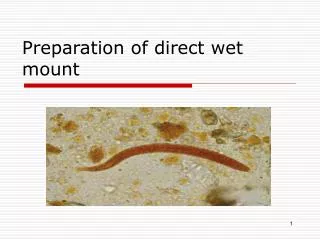
Preparation of direct wet mount. Objectives:. To familiarize the student with the most widely used technique for detection of parasites. To be able to identify the parasite stages (adults, larvae, ova, or cysts). To learn the- students, how to deal with risk samples.

E N D
Objectives: • To familiarize the student with the most widely used technique for detection of parasites. • To be able to identify the parasite stages (adults, larvae, ova, or cysts). • To learn the- students, how to deal with risk samples.
Materials and reagents needed • Microscope, clean slides and coverslip, wooden applicators. • Normal saline solution (9.85% sodium chloride “ Nacl in distilled water). • Pens or markers for labeling
The Microscope • The Microscope is the parasitologist’s main tool. If possible the Microscope- should be binocular; most suitable objectives are the x10, x40, and x100. • The Microscope must be covered and immersion oil removed from the lens -with xylene or ether when not in use. • Calibration of the Microscope Eyepiece Micrometer: • On many occasions measuring the size of suspected parasites in faeces is helpful for identification.(eyepiece micrometer)
Laboratory Methods For Parasites In Faeces • No technique is 100% successful in detecting parasites by a single stool examination, and at least three serial stools must be examined before a patient can be considered free from infections in which stages of parasites would be expected to be found in the faeces. • Whilst clinical symptoms or a case history may provide clues as to which parasites may be present, each faecal specimen should be treated as an unknown, as parasite stages unrelated to the clinical picture may be present.
Faecal specimens • Faecal specimens are examined for the presence of protozoa and helminthes larvae or eggs. • The stages of protozoa found in stools are trophozoites and cysts. The stages of helminthes usually found in stools are eggs and larvae, though whole adult’s worms or segments of worms may also be seen. Adult worms and segments of tapeworms are usually visible to the naked eye, but eggs, larvae, trophozoites, and cysts can be seen only with the microscope.
Collection of faecal specimens 1. Because of the fragile nature of many intestinal parasites, and the need to maintain their morphology for accurate identification, reliable microscopic diagnosis can’t be made unless the stool is collected properly. 2. Approximately 10 gm of fresh faeces uncontaminated by urine, oil, water, dyes or radio-opaque into a clean plastic container. 3. The container should be free from antiseptics and disinfectants. 4. Label all samples clearly with the patient’s name, reference number, date, and time of collection.
Cont. 5. All samples should be accompained by a requisition form from the physician giving relevant clinical details and recent travel history. 6. Samples and forms from patients with a confirmed or suspected diagnosis of certain infectious diseases such as AIDS or hepatitis should be clearly labeled with “Risk of Infection” or “Biohazard” 7. Most viable parasites are susceptible to desiccation or temperature variation. If time lapse between collection and observation is considerable, i.e. more than 4 days, it may be necessary to add some form of preservative to the faeces to retain the morphology as near to the original as possible. 8. Formed samples can be kept in a refrigerator at + 4c for a short while, but not in incubator. 9. Any whole worms or segments passed should be placed in a separate container.
Examination • Macroscopic examination of stool: • As soon as the specimen is received in the laboratory, check: 1.The consistency (degree of moisture) and write one of the following letters on the container: • F (formed), S (soft), L (loose), or W (watery)
2. Abnormal features: • If mucus is present write M, and if blood is present write B. For example, a loose stool with blood and mucus would be recorded as L, B, M. The consistency, or degree of moisture, will be a guide as to whether the trophozoite stage or the cyst stage of protozoa is likely to be present. • If several specimens are received at the same time; those containing blood and mucus should be examined first, followed by liquid specimens. and must be examined within 1 hour after passage. Formed specimens may be examined during the first day.
If no parasites are found • “No ova or parasites seen”, and specify whether this result was obtained by direct examination or by a concentration method (name method used). • Never state categorically: “No parasites”
Microscopic Examination of Wet Mount • Wet mount is the simplest and easiest technique for the examination of faeces, and this method should be performed in all laboratories at the peripheral level. • A wet mount can be prepared directly from faecal material or from concentrated specimens. The basic types of wet mount that should be used for each faecal examination are saline, iodine, and buffered methylene blue.
The saline wet mount • Is used for the initial microscopic examination of stools. It is employed primarily to demonstrate worm's eggs, larvae, protozoan trophozoites, and cysts. • This type of mount can also reveal the presence of red blood cells and white blood cells.
The iodine wet mount • Is used mainly to stain glycogen and the nuclei of cysts, if present. Cysts can usually be specifically identified in this mount. • -The buffered methylene blue (BMB) wet mount should be prepared each time amoebic trophozoites are seen in a saline wet mount, or when their presence is suspected.
Direct saline and iodine mounts 1. With a wax pencil writes the patient’s name or number and the date at the left-hand end of the slide. 2. Place a drop of saline in the center of the left half of the slide and place a drop of iodine solution in the center of the right half of the slide. Note: If the presence of amoebic trophozoites is suspected, warm saline (37c) should be used. 3. With an applicator stick (match or tooth pick), pick up a small portion of the specimen (size of a match head) and mix the drop of saline.
Note • Formed stool: take the portion of stool from an area to include inside and outside parts of the specimen. • Stool with mucus: if mucus is present, label a second slide with the patient’s name or number. Put a drop of saline on the slide, pick up a small portion of mucus and mix with the saline. Trophozoites, if present, are sometimes more readily found in mucus than in the solid parts of the stool. • Loose watery stool: if mucus is not present, pick up a small portion of the stool (any part) and mix with the saline.
Cont. 4. Similarly, pick up a small amount of the stool and mix with the drop of iodine, to prepare an iodine mount. If a wire loop is used, flame it after making the mount. If applicator sticks are used, discard them. 5.Cover the drop of saline and the drop of iodine with a coverslip. Hold the coverslip at an angle, touch the edge of the drop, and lower gently on to the slide. This will reduce the chance of including air bubbles in the mount.
Examination 1. Put the slide with the mounts on the microscope stage and focus on the mount with the x10 or low-power objective. 2. Regulate the light in the microscope field with the sub stage diaphragm. You should be able to see objects in the field distinctly. Too much or too little light is not good. 3. Examine the entire coverslip area with the x10 objective; focus the objective on the top left-hand corner and move the slide systematically backwards and forwards, or up and down.
Cont. 4. When organisms or suspicious material are seen, switch to the high-dry objective, and increase the light by opening the substage diaphragm to observe the detailed morphology. -This is a systematic examination. If mounts are examined in this way, any parasites present will usually be found. If the mount is not examined systematically, parasites may be missed. Examine each microscope field carefully, focusing up and down, before moving to the next field.
Artifacts • Artifacts other things, living or artificial, present in the stool that are not parasites and could mislead the laboratory worker. • Note:“Artifacts not to be mistaken for cysts”.
Artifacts • 1. Blastocystis • 2. Yeasts • 3. Leukocytes • 4. Pus • 5. Coccidia
Plant cells Pollen grains Air bubble Plant fibre Plant hairs Non-human coccidial oocysts Fat droplets Soapy plaques Starch cell Charcot leyden crystals Muscle fibers Fatty acids Macrophage Epithelial cells