Case 2
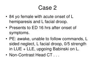
Case 2. 84 yo female with acute onset of L hemiparesis and L facial droop. Presents to ED 16 hrs after onset of symptoms. PE: awake, unable to follow commands, L sided neglect, L facial droop, 0/5 strength in LUE + LLE, upgoing Babinski on L. Non-Contrast Head CT.

Case 2
E N D
Presentation Transcript
Case 2 • 84 yo female with acute onset of L hemiparesis and L facial droop. • Presents to ED 16 hrs after onset of symptoms. • PE: awake, unable to follow commands, L sided neglect, L facial droop, 0/5 strength in LUE + LLE, upgoing Babinski on L. • Non-Contrast Head CT . . .
R frontal wedged-shaped hypodensity c/w infarct; No evidence of hemorrhage
Hyperdense MCA sign (M1 segment) In the M1 segment, thrombus runs parallel to plane of axial CT images, creating the hyperdense MCA sign.
MCA Dot Sign (M2 segment) In M2 and M3 segments, thrombus runs perpendicular to plane of axial CT images, creating the dot sign. Image from: Leary MC et al. Stroke 34 (11) 2636-2640.
MCA Segments (on the insula) Image from: Neafsey E. www.meddean.luc.edu
Differential Dx of Hyperdense MCA • Acute thrombus • Atherosclerotic calcifications (usually bilat) • Elevated Hct (usually bilat) • Herpes simplex encephalitis • subacute stroke 2-5 can be considered potential “false positives”
Clinical Course • Pt admitted, started on Plavix. • Pt remains on the neurology service, with slight improvement of L-sided weakness.
Teaching Points • Primary role of NCHCT in acute stroke pt is to r/o hemorrage. • However, assessing NCHCT for the signs of early ischemic stroke is also possible. • Hyperdense MCA and MCA Dot Signs are very specific findings for MCA occlusion, but have low sensitivity when compared to MCA occlusion demonstrated by angiography: - Sensitivity: 35-40% - Specificity: 95-100% • Prognosis: No CT signs of MCA occlusion > MCA dot sign > hyperdense MCA sign
References • Leary MC, et al. Validation of computed tomographic middle cerebral artery “dot” sign: An angiographic correlation study. Stroke 34 (11): 2636-2640; 2003. • Tomsick T, et al. Thrombus localization with emergency cerebral CT. AJNR 13: 257-263; 1992