Download
1 / 51
540 likes | 916 Views
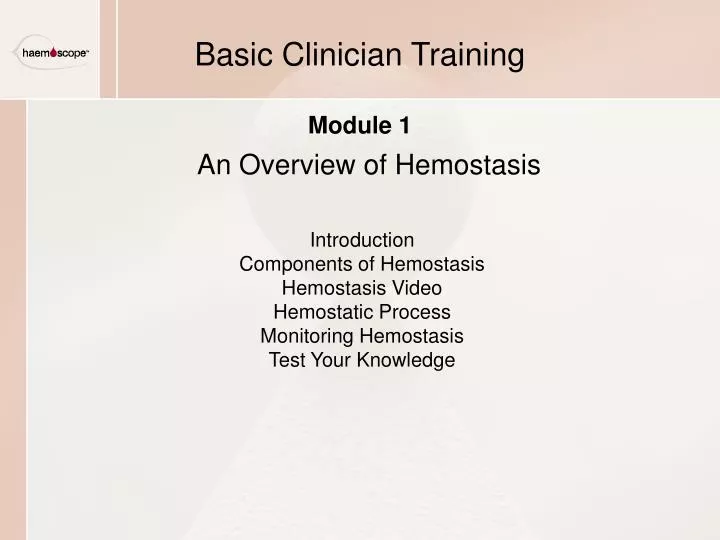
Basic Clinician Training. Module 1. Introduction Components of Hemostasis Hemostasis Video Hemostatic Process Monitoring Hemostasis Test Your Knowledge. An Overview of Hemostasis. Hemostatic System. Introduction Define hemostasis Why monitor hemostasis?. Definition of Hemostasis.

E N D
Basic Clinician Training Module 1 Introduction Components of Hemostasis Hemostasis Video Hemostatic Process Monitoring Hemostasis Test Your Knowledge An Overview of Hemostasis
Hemostatic System Introduction • Define hemostasis • Why monitor hemostasis?
Definition of Hemostasis • Balance between procoagulant and anticoagulant systems • Liquid blood in normal blood vessels • Rapid creation of hemostatic plug at site of injury • Self regulation of complex, dynamic, interactive elements for controlled clot formation and lysis
Administrative Improve patient care Use hemostatic drugs appropriately Lower costs Reduce blood product use Reduce re-operations Reduce thrombotic events Reduce length of stay Clinical Assess risk of bleeding or thrombotic event Personalize hemostatic therapy Monitor efficacy of hemostatic therapy Why Monitor Hemostasis?
The Hemostatic Process:A System Perspective • At least six systems — Proteins • Coagulation pathways • Fibrinolytic pathway • Extra-vascular matrix and tissues — Cells • Platelets • Endothelium • Inflammatory cells • Interdependent components • Self-regulating process Source: Diagnostica Stago
Components of Hemostasis Vascular Vascular Platelets Platelets Interactive Interactive Coagulation Proteins
Intact Endothelium Releases prostacyclin, nitric oxide Expresses heparin-like molecules and thrombomodulin Synthesis and release of tPA Components: Vascular System Endothelium Subendothelium Extra-vascular tissue Platelets Vascular Coagulation Proteins
Components: Vascular SystemEndothelium • Damaged endothelium • Von Willebrand factor • Tissue factor • Fibrinolytic inhibitor (PAI) • Endothelial cells activated by inflammatory mediators • Express tissue factor • Express binding sites for factors IXa and Xa Endothelium Subendothelium Extra-vascular tissue Platelets Vascular Coagulation Proteins
Components: Platelets • Platelets • Normally inactive • Activated by vascular injury • Deform upon activation • Activated platelets Adhesion Activation Secretion Aggregation Procoagulant Activity Platelets Vascular Coagulation Proteins
Components: PlateletsAdhesion • Promoted by collagen • Enhanced by GPIb, vWF Adhesion Activation Secretion Aggregation Procoagulant Activity Platelets Vascular Coagulation Proteins
Components: PlateletsSecretion • Enhances process • Release of dense bodies • Release of α-granules Adhesion Activation Secretion Aggregation Procoagulant Activity Platelets Vascular Coagulation Proteins
Components: PlateletsAggregation • Synthesis and release of thromboxane • Involves GP IIb/IIIa and fibrinogen Adhesion Activation Secretion Aggregation Procoagulant Activity Platelets Vascular Coagulation Proteins
Components: PlateletsThrombin Generation • Activated platelet provides a phospholipid surface • Activation site for coagulation factors, especially V and VIII • Thrombin generation Adhesion Activation Secretion Aggregation Procoagulant Activity Platelets Vascular Coagulation Proteins
Components: Coagulation Proteins • Tissue factor activates factor VII (extrinsic pathway) • Extrinsic and intrinsic pathways • Converge at factor X • Models well in vitro • Do not model well in vivo • Extrinsic initiates thrombin generation • Intrinsic amplifies thrombin generation (factor V and VIII) Platelets Vascular Coagulation Proteins Extrinsic and Intrinsic Pathways
Components: Coagulation ProteinsThrombin Generation • Thrombin generation • Pivotal point for coagulation (PT, aPTT measure only 5% of total thrombin production) • Self promoting (activates Factor XI) • Self limiting (thrombin activates thrombomodulin) Platelets Vascular Thrombin Coagulation Proteins Extrinsic and Intrinsic Pathways
Cascade Model Area of Injury Change in Platelet Shape Endothelial Cells Collagen Platelet AA ADP CoagulationCascade tPA Fibrinolysis Fibrin Strands Plasminogen Plasmin Degradation Products
Subendothelial cells and leukocytes Express tissue factor Provide reaction surface for coagulation protein activation and binding (TF/VIIa complex formed) Lead to thrombin generation (Factor X, IX, VIII, V, and XI) Phospholipid surface Components: Cellular Elements
Cell-Based Model Tissue factor bearing cells • Reflects in vivo • Occurring on cell surfaces • Tissue factor bearing cells • Platelets • Overlapping phases: • Initiation (TF bearing cells) • Amplification (platelets) • Propagation (platelets) • The coagulation cascades are still important, but are cell-based • extrinsic pathway: surface of tissue factor bearing cells • intrinsic pathway: surface of platelets • Routine coagulation tests do not represent the cell-based model of hemostasis 1. Initiation IIa 2. Amplification Platelets 3. Propagation IIa Activated platelets [Monroe, DM. et al. Arterioscler Thromb Vasc Biol. 2002;22:1381]
Normal/Balanced Hemostasis • Multiple feedback mechanisms maintain balance • Balance is maintained © 2005 Haemoscope Corporation
Abnormal/Unbalanced Hemostasis • Imbalance when mechanisms are overwhelmed • Surgery • Trauma • Disease • Drugs Hypocoagulable Hypercoagulable
Hemostasis Video This version does not contain a video. Your local representative may be able to provide an updated version.
Hemostasis • Clot: The end product of hemostasis • Platelet plug formation (white clot) • Platelet-fibrin clot formation (red clot) • Fibrinolysis
Platelet Plug Formation : exposure to collagen • Endothelial damage • Promotes platelet adherence and activation • Platelet recruitment • Platelet aggregation • Results in formation of platelet plug (white clot)
Initiation of Thrombin Generation Endothelial damage Exposure to tissue factor Initiation of extrinsic pathway Initiation of thrombin generation Platelet Activation Intrinsic pathway Amplification of thrombin generation
Fibrin-Platelet Clot Formation • Thrombin generation the pivotal point of the coagulation process • Thrombin prothrombotic actions • Platelet activation • Amplification of thrombin generation • Fibrin clot development through conversion of fibrinogen to fibrin • Result: fibrin-platelet clot (red clot)
Fibrin Formation:Initiation of Fibrinolysis • Tissue plasminogen activator binds to fibrin • Converts plasminogen to plasmin • Plasmin breaks down fibrin tPA Fibrin Stands Plasminogen Plasmin Degradation Products
Hemostasis Monitoring Hemostasis
Cascade Model: Tests Endothelial Cells Change in Platelet Shape Area of Injury • Represents hemostasis • Two independent activation pathways • Pathways converge at the final common pathway • PT, aPTT: based on cascade model • Measure coagulation factor interaction in solution • Determine if adequate levels of coagulation factors are present for clot formation Collagen White Clot Platelet AA ADP Thrombin Generation CoagulationCascade PT aPTT Platelet counts Red Clot tPA Fibrin Strands Fibrinolysis Plasminogen Plasmin Degradation Products
Monitoring: Cell Based Model • Whole blood sample • Platelets • Coagulation factors • Cellular/plasmatic factors • TEG® analysis • Coagulation factors • Fibrinogen • Platelets • Fibrinolytic factors • Inflammatory cells • Mediators
Monitoring: Hemostatic Process • Hemostatic process: cell based model plus red blood cells, white blood cells, etc. • Activation clot formation clot lysis • Entire process: TEG system
Monitoring Insights • Results are used in conjunction with patient status: • Patient clinical condition (bleeding/not bleeding) • Phase in medical intervention • Type and dose of drug therapy • Patient history • TEG testing shows net effect “whole picture” of hemostasis at that point in time: • Identifies a “factor deficiency,” but not which factor • Identifies a platelet defect, but does not distinguish between platelet deficiency and platelet dysfunction
Monitoring Optimization • Trend analysis • Hemostatic state over time • Individual patient analysis • Inhibitor effects
Summary • Hemostasis • Interactive components • Balance • Hemostatic tests • Cascade model: limited (PT, aPTT) • Cell-based model: whole blood (TEG) • Monitoring hemostasis • Appropriate drugs • Reduction in health care costs • Personalized treatment improved patient care
Basic Clinician Training Test your knowledge of hemostasis by answering the questions in the slides that follow. Hemostasis Hemostatic Monitoring
Exercise 1 Normal hemostasis is characterized by a functional ______ between the procoagulant pathways/components and the antithrombotic and anticoagulant pathways/components. Answer: page 43
Exercise 2 What is the typical initiating event of the hemostatic process? • Platelet activation • Thrombin generation • Endothelial damage • Plasmin generation Answer: page 44
Exercise 3 What is the pivotal point in the activation of the coagulation pathways? • Tissue factor expression • FXII activation • FXa generation • Thrombin generation • Fibrin formation • Platelet activation Answer: page 45
Exercise 4 Which coagulation pathway is responsible for the initiation of thrombin generation? a) Intrinsic b) Extrinsic Which coagulation pathway is responsible for the amplification of thrombin generation? a) Intrinsic b) Extrinsic Answer: page 46
Exercise 5 In the cell-based model of hemostasis, where do the intrinsic and extrinsic pathway activities occur? a) On neutrophils b) On the tissue-factor bearing cells and platelets c) In the plasma d) On endothelial cells Answer: page 47
Exercise 6 Which of the following statements does not describe PT and aPTT tests? • They both measure how coagulation factors interact in solution. • They both use fibrin formation as a static end point. • They both demonstrate the effect of thrombin generation on platelet function. • They both demonstrate the function of the extrinsic and intrinsic pathways, respectively. Answer: page 48
Exercise 7 The TEG system is a whole blood hemostasis analyzer that can measure the contribution of which of the following hemostatic components? (select all that apply) • Enzymatic factor • Fibrinogen • Platelets • Fibrinolytic pathway • Endothelial cells Answer: page 49
Exercise 8 The TEG analyzer provides results that help distinguish between surgical bleeding and bleeding due to a coagulopathy. True or False? Answer: page 50
Answer to Exercise 1 Normal hemostasis is characterized by a functional balancebetween the procoagulant pathways/components and the antithrombotic and anticoagulant pathways/components.
Answer to Exercise 2 What is the typical initiating event of the hemostatic process? a) Platelet activation b) Thrombin generation c) Endothelial damage d) Plasmin generation
Answer to Exercise 3 What is the pivotal point in the activation of the coagulation pathways? a) Tissue factor expression b) FXII activation c) FXa generation d) Thrombin generation e) Fibrin formation f) Platelet activation
Answer to Exercise 4 Which coagulation pathway is responsible for the initiation of thrombin generation? a) Intrinsic b) Extrinsic Which coagulation pathway is responsible for the amplification of thrombin generation? a) Intrinsic b) Extrinsic
Answer to Exercise 5 In the cell-based model of hemostasis, where do the intrinsic and extrinsic pathway activities occur? a) On neutrophils b) On the tissue-factor bearing cells and platelets c) In the plasma d) On endothelial cells
Answer to Exercise 6 Which of the following statements does not describe PT and aPTT tests? a) They both measure how coagulation factors interact in solution b) They both use fibrin formation as a static end point c) They both demonstrate the effect of thrombin generation on platelet function d) They both demonstrate the function of the extrinsic and intrinsic pathways, respectively.
Answer to Exercise 7 The TEG system is a whole blood hemostasis analyzer that can measure the contribution of which of the following hemostatic components? (select all that apply) a) Enzymatic factor b) Fibrinogen c)Platelets d)Fibrinolytic pathway e) Endothelial cells
Answer to Exercise 8 The TEG analyzer provides results that help distinguish between surgical bleeding and bleeding due to a coagulopathy. True