Download
1 / 18
180 likes | 222 Views
Explore the mechanical properties of tendons and ligaments, stress-strain curves, ligament injuries classification, management strategies, tendon injuries, and tendon pathologies such as tendonitis vs. tendinopathy.

E N D
Mechanical Behavior of Tendon & Ligament Physiotherapy Semester – I
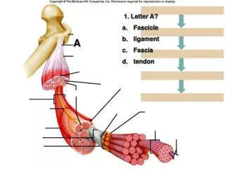
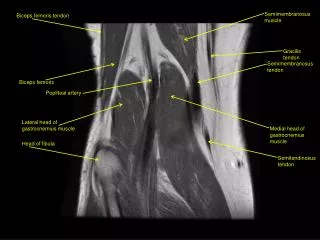
Mechanical Behavior of Tendons and Ligaments Tendons Strong Viscoelastic structures Sustain the high tensile forces Sufficiently flexible to angulate around bone surfaces Ligaments Pliant and flexible to allow natural movements of the bones Strong & inextensible to offer suitable resistance to applied forces
Load-elongationResponse 1: Primary or "Toe" region … The tissue elongated easily with a small increase in load 2: Secondary or "Linear" region …. The fibers straightened out & fibers’ stiffness increased Greater force is required for elongation Deformation of the tissue began 3: End of Secondary Region (P lin.)…. Progressive failure of the collagen fibers Irreversible deformation 4: Maximum Load (Pmax) ….. Complete failure occurred rapidly and Lost its ability to support loads
Load-elongation Curves Load-elongation Curve of tendon Load-elongation Curve of lig. flavum
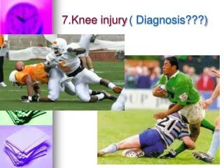
Clinical Consideration Ligament Failure ……. Progressive failure against increasing load Stress – Strain Relationship Microfailure & gross failure of ligament Joint displaced several millimeter Damage to the surrounding tissues as well e.g. : ACL Injury in soccer as a result of an abnormal torque in rotation of the knee
Load-displacement curve of ACL Injury Load imposed on the ACL during the ant. drawer test Load placed on the ligament during physiological activity Load imposed on the ligament from partial injury to complete rupture (Microfailure region) Note: Why ACL Injury can lead to EARLY osteoarthritis? Load-displacement curve
Ligament Injuries are classified clinically into three Categories Grade-I Sprain ….. Mild pain, No joint instability, Micro-tear of few fibers Grade-II Sprain ….. Severe pain, Some joint instability, Partial ligament rupture (Progressive failure of the collagen fibers) 50% decrease in Ligament strength (increase laxity with a definite end point) joint stability is tested usually under anesthesia Grade-III Sprain …. Severe pain during trauma but less pain after injury Gross instability (excessive joint laxity with no firm end point) Complete ruptured but a few may still be intact
Management of Ligament Sprains Grade I & II Sprain …. First aid Management elecrophysical agents, joint mobilization & soft tissue massage muscle strengthening, proprioceptive training & Functional training Grade III Sprain …. First aid Managementsurgery ( repair or reconstruction) / protective bracing muscle strengthening, proprioceptive training & Functional training
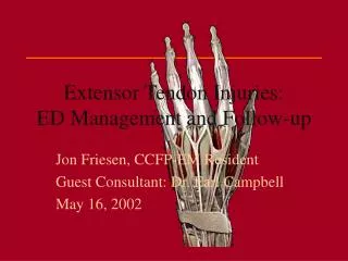
Clinical Consideration of Tendon Injuries Two additional factors become important in tendons …… The amount of force produced by muscle contraction The cross-sectional area of the tendon Muscle contraction exerts a tensile stress on the tendon. rapid eccentric contraction of the muscle increases the stress Tensile strength of a tendon α Cross-sectional area of the tendon Why muscle ruptures are more common than are ruptures through a tendon?
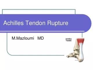
Load Deformation Curve Of Achilles Tendon Rupture First region ….. a normal physiological toe-loading response Linear region …. high load is producing a higher deformation within the tendon structure Overuse region …. Repetitive higher strains with insufficient time for the healing process
Tendonitis vs. Tendinopathy Tendonitis …… inflammation of the tendon itself Tendinopathy …. The primary pathology is Collagen degeneration Histological examination reveals separation of collagen bundles, increased hydrophilic ground substances, increased poor quality blood vessels ( neovascularization), and absent inflammatory cells Common sites for tendinopathies are rotator cuff, common extensors origion of elbow, extensor carpi redialis brevis, patellar and Achilles tendons Other sites add. Longus, biceps, tibialis post. And flex. Hallucis longus tendons.
Grading of Tendinopathy Descriptive Grading of Tendinopathy …. Mild Tendinopathy…. Pain after activity only / no pain during activity Moderate Tendinopathy …. Pain with sporting activity but not with ADL Severe Tendinopathy …. Pain during ADL Tendon-specific scoring system….. The VISA Score e.g. patellar tendinopathy
Tendinopathy Characteristic features ….. Pain some time after exercise or, more frequently, the following morning when the athlete rises Can ‘run through’ the pain or pain disappears in warming up & return after exercise when cooling down On Examination: Local tenderness, thickening, swelling, crepitus Management …. Eccentric loading exercises vs. concentric exercise Surgery is indicated on clinical basis ( stripping of paratenon, release of adhesions, removal of degenerative tissues, repair of a partial tear or excision of a torn flap)
Age- related changes of collagen Collagen cross-links vs. Aging: The greater the no. & quality of cross-links, the more tensile strength of collagen (Viidik, Danielsen, & Oxlund, 1982) Collagen fibril diameter vs. Aging: Fibril diameter αthetensile strength of ligament and tendons Fibril diameter in adults (20-60 years) & in elderly (>60 years) decreases remarkably (Strocchi et al., 1996) Water content & concentration of collagen vs. Aging: The water content & collagen conc. decreases significantly in ligament as age progresses after maturation (Amiel, 1991). So,tensile strength & stiffness of the ligament & tendon decreases
Mobility vs. Mechanical behavior of Ligament & Tendon Functional adaptation of ligament & tendon in response to the mechanical demands placed on them The ligaments are more stronger & stiffer when subjected to increasing stress and more weaker & lesser stiffer when subjected to decreasing stress(Noyes et al., 1977a) Physical training increases the tensile strength of tendons and ligament-bone interface(Woo et al., 1981). Research study of medial collateral ligament strength: The ligaments of the exercised dogs for 6 weeks were stronger & stiffer than those of the control dogs & the collagen fiber bundles had larger diameters(Tipton and coworkers, 1970)
Diabetes Mellitus Diabetes mellitus VS. Musculoskeletal disorders Diabetics vs. Nondiabetics …. Higher rates of tendon contracture (29 vs. 9%) Tenosynovitis (59 vs. 7%) Joint stiffness (40 vs. 9%) Capsulitis (16 vs. 1%) Also causes osteoporosis (Carvallo et al., 1991; Lancaster et al., 1994) The tissue elastic properties did not differ between the diabetic and the control group. The viscous component of the tissue response, however, was increased in the hyperglycemic group. Insulin therapy seems to lessen such alterations (Duquette, 1996)
Nonsteroidal Anti-inflammatory Drugs NSAIDs (aspirin, acetaminophen and indomethacin) are frequently used in painful conditions of the musculoskeletal system also widely used in the treatment of soft tissue injuries such as inflammatory disorders and partial ruptures of tendons and ligaments Vogel (1977) found that treatment with indomethacin resulted in increased tensile strength in rat tail tendons. An increase in the proportion of insoluble collagen and in the total collagen content also was observed Ohkawa (1982) found increased tensile strength in the periodontium of rats after indomethacin treatment Carlstedt and associates (1986a, 1986b) found that indomethacin treatment increased the tensile strength in developing and healing plantaris longus tendons in the rabbit and noted that the mechanism for this increase was probably an increased cross-linkage of collagen molecules