Glomerular Diseases: Pathology, Causes, and Syndromes
560 likes | 580 Views
Explore primary and systemic glomerulopathies, including causes, immune complex types, and clinical syndromes. Learn about renal functions, disease morphology, and diagnostic approaches in glomerulonephritis.

Glomerular Diseases: Pathology, Causes, and Syndromes
E N D
Presentation Transcript
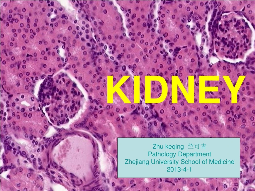
The Kidney KIDNEY Zhu keqing 竺可青 Pathology Department Zhejiang University School of Medicine 2013-4-1
Glomerular Diseases Primary Glomerulopathies 原发性 • Acute diffuse proliferative glomerulonephritis • Poststreptococcal • Non-poststreptococcal • Rapidly progressive (crescentic) glomerulonephritis • Membranous glomerulopathy • Minimal change disease • Focal segmental glomerulosclerosis • Membranoproliferative glomerulonephritis • IgA nephropathy • Chronic glomerulonephritis
Glomerular Diseases Systemic Diseases with Glomerular Involvement 继发性 • Systemic lupus erythematosus • Diabetes mellitus • Amyloidosis • Goodpasture syndrome • Microscopic polyarteritis/polyangiitis • Wegener granulomatosis • Henoch-Schönlein purpura • Bacterial endocarditis Hereditary Disorders • Alport syndrome • Thin basement membrane disease • Fabry disease
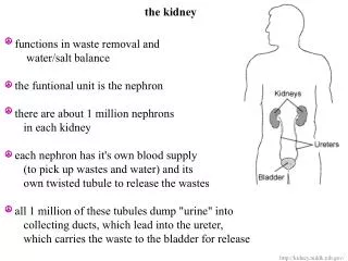
肾小球组织学 • 肾小球由毛细血管丛及肾球囊构成,前者是毛细血管袢,后者壁薄衬以单层扁平上皮(脏层与壁层上皮;二者间为腔。) 肾小球的超微结构 • 肾小球囊的脏层存在肾小球滤过膜,有三层组织结构, 包含 • 内侧为连续性不强的内皮细胞层; • 中间是基膜,是粘多糖形成的网架; • 外侧为多数足突附着于基膜,其间见甚薄的空隙,称滤过隙。 • 足突是脏层上皮(足细胞)的最后胞突分枝。 • 血管袢的中央有系膜细胞及其基质。
病因 • 肾小球肾炎简称肾炎,是由于各种免疫复合物引起的两侧肾脏肾小球弥漫性增生性炎,属Ⅲ型变态反应,即免疫复合物肾炎。 (一)抗原 • 1.内源性抗原 包括肾小球性(肾小球基底膜抗原、足细胞的足突抗原、内皮细胞和系膜细胞的细胞膜抗原等)和非肾小球性(DNA、核抗原、免疫球蛋白、肿瘤抗原等)。 • 2.外源性抗原 生物性病原体,最常见是链球菌感染后的产物,肝炎病毒、疟疾以及药物、异种血清也可成为抗原。 (二)免疫复合物类型 • 肾小球原位免疫复合物 占10%。肾小球基膜内固有的或植入的抗原成分与循环中的抗体结合,形成复合物。免疫荧光检查呈光滑线形荧光。 • 循环免疫复合物 占90%。由外源性抗原或非肾小球性内源性抗原和相应的抗体结合,随血流经肾小球时沉积于局部。免疫荧光检查呈不连续的颗粒状荧光。
发病机理 • 免疫复合物激活补体,进而激活白细胞,白细胞释放氧自由基,蛋白酶及花生四烯酸等,引起肾小球的损伤,导致增生性渗出性炎. • 免疫复合物通过作用于血小板,系膜细胞及单核细胞,使细胞因子产生增加, 损伤肾小球,导致增生性渗出性炎.
Various types of glomerulonephritis are characterized by one or more of four basic tissue reactions基本病理变化 Hypercellularity.增生性炎症 • Cellular proliferation of mesangial or endothelial cells • Leukocytic infiltration, consisting of neutrophils, monocytes, and, in some diseases, lymphocytes • Formation of crescents. Basement Membrane Thickening. Hyalinization and Sclerosis.
Clinical syndrome • Acute nephritic syndrome • Rapidly progressive nephritic syndrome • Nephrotic syndrome • Asymptomatic hematuria or proteinuria • Chronic nephritic syndrome • Acute renal failure • Chronic renal failure • Renal tubular defects • Urinary tract infection • Nephrolithiasis (renal stone) • Urinary tract obstruction and renal tumors
The Glomerular Syndromes 1. Acute nephritic Syndrome Hematuria, azotemia, variable proteinuria, oliguria, edema, and hypertension 2. Rapidly progressive glomerulonephritis Acute nephritis, proteinuria, and acute renal failure 3. Nephrotic syndrome >3.5 gm proteinuria, hypoalbuminemia,hyperlipidemia, lipiduria 4. Chronic renal failure Azotemia → uremia progressing foryears 5. Asymptomatic hematuria or proteinuria Glomerular hematuria; subnephrotic proteinuria
1Acute Proliferative (Poststreptococcal, Postinfectious) Glomerulonephritis Morphology. • The classic diagnostic picture is one of enlarged, hypercellular glomeruli大红肾/蚤咬肾. The hypercellularity is caused by • (1) infiltration by leukocytes, both neutrophils and monocytes; • (2) proliferation of endothelial and mesangial cells; and • (3) in severe cases by crescent formation. • By immunofluorescence microscopy, there are granular deposits of IgG, IgM, and C3 in the mesangium and along the basement membrane. Although almost universally present, they are often focal and sparse. • The characteristic electron microscopic findings are discrete, amorphous, electron-dense deposits on the epithelial side of the membrane, often having the appearance of "humps", presumably representing the antigen- antibody complexes at the epithelial cell surface. Subendothelial and intramembranous deposits are also commonly seen, and mesangial deposits may be present.
Acute Proliferative (Poststreptococcal, Postinfectious) Glomerulonephritis • More than 95% of affected children eventually recover totally with conservative therapy aimed at maintaining sodium and water balance. 儿童预后好 • A small minority of children (perhaps less than 1%) do not improve, become severely oliguric, and develop a rapidly progressive form of glomerulonephritis. • Some of the remaining patients may undergo slow progression to chronic glomerulonephritis with or without recurrence of an active nephritic picture. • Prolonged and persistent heavy proteinuria and abnormal GFR mark patients with an unfavorable prognosis. 持续蛋白尿预后差 • In adults, the disease is less benign.成人预后差 • Although the overall prognosis in epidemics is good, in only about 60% of sporadic cases do the patients recover promptly. • In the remainder, the glomerular lesions fail to resolve quickly, as manifested by persistent proteinuria, hematuria, and hypertension. In some of these patients, the lesions eventually clear totally, but others develop chronic glomerulonephritis. Some patients will develop a syndrome of rapidly progressive glomerulonephritis.
急性弥漫性毛细血管内增生性肾小球肾炎(小结)急性弥漫性毛细血管内增生性肾小球肾炎(小结) • Diffuse endocapillary proliferativeglomerulonephritis 简称急性肾炎,很常见,小儿多,病理属急性增生性炎症,属变态反应。大多数与溶血性链菌感染有关,故又称链球菌感染后肾炎。 • 1.病理变化 光镜: • 增生性病变:双侧肾脏几乎全部肾小球的体积增大,细胞核数目增多(密集,深染)致使毛细血管腔狭窄。 • 渗出性病变:有少许中性白细胞、少量纤维素渗出及红细胞(有时红细胞甚多)漏出到球囊腔内。 • 变质性病变:肾小管浊肿 肉眼:大红肾,蚤咬肾。 电镜:见血管间质细胞及内皮细胞增生;足突下基膜上有散在的驼峰样免疫复合物沉积。 • 2.临床病理联系 • 急性肾炎综合征 • 少尿、无尿 • 蛋白尿、管型尿、血尿 • 水肿 • 高血压 • 3.结果 • 95%以上患者治愈,少数转为慢性,极少数发展为快速进行性肾炎。
2 RAPIDLY PROGRESSIVE (CRESCENTIC) GLOMERULONEPHRITIS Rapidly progressive glomerulonephritis (RPGN) is a syndrome associated with severe glomerular injury and does not denote a specific etiologic form of glomerulonephritis. It is characterized clinically by rapid and progressive loss of renal function associated with severe oliguria and (if untreated) death from renal failure within weeks to months. Regardless of the cause, the classic histologic picture is characterized by the presence of crescents in most of the glomeruli (crescentic glomerulonephritis). These are produced in part by proliferation of the parietal epithelial cells lining Bowman capsule and in part by infiltration of monocytes and macrophages.
Rapidly Progressive Glomerulonephritis (RPGN) Type I RPGN (Anti-GBM Antibody) • Idiopathic • Goodpasture syndrome Type II RPGN (Immune Complex) • Idiopathic • Postinfectious • Systemic lupus erythematosus • Henoch-Schönlein purpura (IgA) • Others Type III RPGN (Pauci-Immune) • ANCA associated • Idiopathic • Wegener granulomatosis • Microscopic polyarteritis nodosa/microscopic polyangiitis
Rapidly Progressive Glomerulonephritis (RPGN) Morphology. • The kidneys are enlarged and pale, often with petechial hemorrhages on the cortical surfaces. 大白肾 • The histologic picture, however, is dominated by the formation of distinctive crescents. Crescents are formed by proliferation of parietal cells and by migration of monocytes and macrophages into the urinary space. • Electron microscopy may, as expected, disclose subepithelial deposits in some cases, but in many cases, it shows distinct ruptures in the GBM, the severe injury that allows leukocytes, proteins, and inflammatory mediators into the urinary space, where they trigger the crescent formation. • By immunofluorescence microscopy, postinfectious cases exhibit granular immune deposits; Goodpasture syndrome cases show linear fluorescence for immunoglobulin and complement.
Clinical Course. • In Goodpasture syndrome, the course may be dominated by recurrent hemoptysis or even life-threatening pulmonary hemorrhage. Serum analyses for anti-GBM antibodies, antinuclear antibodies, and ANCA are helpful in the diagnosis of specific subtypes. • Although milder forms of glomerular injury may subside, the renal involvement is usually progressive over a matter of weeks and culminates in severe oliguria. • Recovery of renal function may follow early intensive plasmapheresis 血透(plasma exchange) combined with steroids and cytotoxic agents in Goodpasture syndrome. • Other forms of RPGN also respond well to steroids and cytotoxic agents. • Despite therapy, patients may eventually require chronic dialysis or transplantation.
快速进行性肾小球肾炎(小结) • 又称弥漫性新月体性肾小球肾炎(diffuse crecsentic glomerulonephritis)。临床经过发展迅速,常因少尿无尿、肾功能进行性障碍性在短期内(数周或数月)死亡。25%由急性肾炎转化而来,25%合并抗基底膜肾炎,50%原因未明。 • 1.病理变化 • 光镜: • 肾小球囊壁层上皮增生,与与渗出的单核细胞形成新月体(细胞性新月体),随后纤维化并与毛细血管袢粘连(纤维性新月体),致肾小球快速纤维化、玻璃样变。阻塞尿液出口。 • 肉眼:大白肾 • 电镜:一些病例有免疫复合物沉积,全部病例发现有基底膜局灶性破裂。 • 2.临床联系 • 快速进行性肾炎综合征: • 少尿、无尿。 • 血尿、蛋白尿严重。 • 快速进行性肾功能衰竭。 • 3.结果 • (1)预后极差,80%病例在半年内死亡。 • 预后取决新月体出现数量:50%新月体者预后较好;50-80%新月体者发展缓慢;80-90%新月体者死亡。 • (2)转为慢性硬化性肾小球肾炎
3 The manifestations of the nephrotic syndrome include: • Massive proteinuria, with the daily loss of 3.5 gm or more of protein (less in children) • Hypoalbuminemia, with plasma albumin levels less than 3 gm/dL • Generalized edema • Hyperlipidemia and lipiduria
Causes of Nephrotic Syndrome Prevalence (%) Children Adults Primary Glomerular Disease • Membranous glomerulopathy 5 30 • Minimal change disease 65 10 • Focal segmental glomerulosclerosis 10 35 • Membranoproliferative glomerulonephritides 10 10 • Other proliferative glomerulonephritis (focal,"pure mesangial," IgA nephropathy) 10 15 Systemic Diseases • Diabetes mellitus • Amyloidosis • Systemic lupus erythematosus • Drugs (nonsteroidal anti-inflammatory, penicillamine , "street heroin") • Infections (malaria, syphilis, hepatitis B and C, acquired immunodeficiency syndrome) • Malignant disease (carcinoma, lymphoma) • Miscellaneous (bee-sting allergy, hereditary nephritis)
1 MEMBRANOUS GLOMERULOPATHY (MEMBRANOUS NEPHROPATHY) • Membranous glomerulopathy is the most common cause of the nephrotic syndrome in adults. • It is characterized by diffuse thickening of the glomerular capillary wall and the accumulation of electron-dense, immunoglobulin-containing deposits along the subepithelial side of the basement membrane.
Membranous glomerulopathy • The most notable such associations are as follows: • Drugs (penicillamine , captopril , gold, nonsteroidal anti-inflammatory drugs [NSAIDs]): 1% to 7% of patients with rheumatoid arthritis treated with penicillamine or gold (drugs now used infrequently for this purpose) develop membranous glomerulopathy. • Underlying malignant tumors, particularly carcinoma of the lung and colon and melanoma. According to some investigators, these are present in up to 5% to 10% of adults with membranous glomerulopathy. • SLE. About 15% of glomerulonephritis in SLE is of the membranous type. • Infections (chronic hepatitis B, hepatitis C, syphilis, schistosomiasis, malaria) • Other autoimmune disorders, such as thyroiditis • In about 85% of patients, no associated condition can be uncovered, and the disease is considered idiopathic.
Clinical Course of Membranous glomerulonephritis • In a previously healthy individual, this disorder usually begins with the insidious onset of the nephrotic syndrome or, in 15% of patients, with non-nephrotic proteinuria. Hematuria and mild hypertension are present in 15% to 35% of cases. • It is necessary in any patient to first rule out the secondary causes described earlier, since treatment of the underlying condition (malignant neoplasm, infection, or SLE) or discontinuance of the offending drug can reverse progression.
The course of the disease is variable but generally indolent. In contrast to minimal change disease, the proteinuria is nonselective and does not usually respond well to corticosteroid therapy. • Although proteinuria persists in more than 60% of patients, only about 10% die or progress to renal failure within 10 years, and no more than 40% eventually develop renal insufficiency. • Concurrent sclerosis of glomeruli in the renal biopsy at the time of diagnosis is a predictor of worse prognosis.
弥漫性膜性肾小球肾炎(小结) • 弥漫性膜性肾小球肾炎(diffuse membranous glomerulonephritis)简称膜性肾炎,认为是早期性肾炎,进展较慢。 • 1.病理改变 • 光镜:弥漫性肾小球血管基底膜增厚(白金耳样) • 肉眼:大白肾 • 电镜:“屋顶+大钉”,即大量屋顶形沉积物加上基膜物质向外刺状增生。二者结合基膜因而增厚。晚期更厚,虫蚀样。 • 2.临床病理联系 • 典型肾病综合症:高度蛋白尿。高度水肿。高脂血症和脂尿。低血浆白蛋白 • 3.结果 病程发展较慢,大多数病例晚期肾小球纤维化,40%病例死于肾衰。一些病例可治愈。
2 MINIMAL CHANGE DISEASE (LIPOID NEPHROSIS) • This relatively benign disorder is the most frequent cause of nephrotic syndrome in children, but it is less common in adults. 儿童多 • It is characterized by diffuse effacement of foot processes of epithelial cells in glomeruli that appear virtually normal by light microscopy.弥漫性上皮细胞足突消失 • The peak incidence is between 2 and 6 years of age. The disease sometimes follows a respiratory infection or routine prophylactic 预防immunization. • Its most characteristic feature is its usually dramatic response to corticosteroid therapy.
MINIMAL CHANGE DISEASE (LIPOID NEPHROSIS) Etiology and Pathogenesis • Several features of the disease point to an immunologic basis,including • (1) the clinical association with respiratory infections and prophylactic immunization; • (2) the response to corticosteroids and/or other immunosuppressive therapy; (3) the association with other atopic遗传过敏disorders (e.g., eczema湿疹, rhinitis鼻炎); • (4) the increased prevalence of certain HLA haplotypes in patients with minimal change disease ; • (5) the increased incidence of minimal change disease in patients with Hodgkin disease, in whom defects in T cell-mediated immunity are well recognized.
Morphology. • The glomeruli are normal by light microscopy. By electron microscopy, the basement membrane appears normal, and no electron-dense material is deposited. • The principal lesion is in the visceral epithelial cells, which show auniform and diffuse effacement of foot processes足突病。 • The visceral epithelial changes are completely reversible after corticosteroid therapy, concomitant with remission免除of the proteinuria. • The cells of the proximal tubules are often laden with lipid and protein, reflecting tubular reabsorption of lipoproteins passing through diseased glomeruli (thus, the historical term lipoid nephrosis). 脂性肾病 • Immunofluorescence studies show no immunoglobulin or complement deposits.
Clinical Course • Despite massive proteinuria, renal function remains good, and there is commonly no hypertension or hematuria. The proteinuria usually is highly selective, most of the protein consisting of albumin. • Most children (more than 90%) with minimal change disease respond rapidly to corticosteroid therapy. However, the nephrotic phase may recur, and some patients may become steroid dependent or resistant. • Nevertheless, the long-term prognosis for patients is excellent, and even steroid-dependent disease resolves when children reach puberty. 青春期 • Although adults are slower to respond, the long-term prognosis is also excellent.预后好
轻微病变性肾小球肾炎(小结) • 轻微病变性肾小球肾炎(minimal change glomerulonephritis)又称脂性肾病(lipoid nephrosis)。 • 1.病理变化 • 肉眼:大白肾 • 光镜:肾小球无明显改变,肾小管脂肪变。 • 电镜:肾小球仅见足突融合,无沉积物或其它改变。 • 2.临床联系: • 肾病综合症:高蛋白尿。高度水肿。高脂血症和脂尿。低血浆白蛋白 • 3.结果 • 本病多见小儿,用可的松治疗,90%患儿可被治愈。
3 FOCAL SEGMENTAL GLOMERULOSCLEROSIS • As the name implies, this lesion is characterized by sclerosis of some, but not all, glomeruli (thus, it is focal); and in the affected glomeruli, only a portion of the capillary tuft is involved (thus, it is segmental). • Focal segmental glomerulosclerosis is frequently accompanied clinically by the nephrotic syndrome or heavy proteinuria.
FOCAL SEGMENTAL GLOMERULOSCLEROSIS Morphology • By light microscopy, the segmental lesions may involve only a minority of the glomeruli and may be missed if the biopsy specimen contains an insufficient number of glomeruli. Lipid droplets and foam cells are often present. • On electron microscopy, both sclerotic and nonsclerotic areas show the diffuse effacement of foot processes characteristic of minimal change disease, but in addition, there may be focal detachment of the epithelial cells with denudation剥脱of the underlying GBM. • By immunofluorescence microscopy, IgM and C3 may be present in the sclerotic areas and/or in the mesangium. In time, this leads to total sclerosis of glomeruli, with pronounced tubular atrophy and interstitial fibrosis.
Clinical Course. • There is little tendency for spontaneous remission in idiopathic focal segmental glomerulosclerosis, and responses to corticosteroid therapy are variable. • In general, children have a better prognosis than adults do. • Progression of renal failure occurs at variable rates. About 20% of patients follow an unusually rapid course, with intractable难治massive proteinuria ending in renal failure within 2 years. • Recurrences are seen in 25% to 50% of patients receiving allografts.
局灶性节段性肾小球硬化(小结)(focal segmental glomeralosclerosis) • 局灶性:仅累及少数或部分肾小球。 • 节段性:仅累及肾小球的部分血管丛。 • 病变特点: • 早期仅少数肾小球或肾小球的部分毛细血管丛受累(萎陷、系膜增宽、硬化、玻璃样变)。 • 病变继续发展,受累的肾小球逐渐增多。 • 有些肾小球毛细血管丛可全部纤维化、硬化,最终可发展为终期肾。
4 MEMBRANOPROLIFERATIVE GLOMERULONEPHRITIS • Membranoproliferative glomerulonephritis (MPGN) is characterized histologically by alterations in the basement membrane, proliferation of glomerular cells, and leukocyte infiltration. • Because the proliferation is predominantly in the mesangium, a frequently used synonym is mesangiocapillary glomerulonephritis. 系膜增生 • MPGN accounts for 10% to 20% of cases of nephrotic syndrome in children and young adults. Some patients present only with hematuria or proteinuria in the non-nephrotic range, and others have a combined nephrotic-nephritic picture. • Like many other glomerulonephritides, MPGN either can be associated with other systemic disorders and known etiologic agents (secondary MPGN) or may be idiopathic (primary MPGN).
Morphology • By light microscopy, the glomeruli are large and hypercellular. The hypercellularity is produced both by proliferation of cells in the mesangium and so-called endocapillary cell proliferation involving capillary endothelium and infiltrating leukocytes.系膜增生 • Within the besement membrane there is inclusion or interposition of cellular elements, which can be of mesangial, endothelial, or leukocytic origin. Such interposition gives rise to the appearance of “split” basement membranes.系膜插入 • The glomerular capillary wall often shows a “double-contour” or “tram-track” appearance, especially evident in silver or PAS stains. This is caused by “duplication” of the basement membrane, usually as the result of new basement membrane synthesis. 双轨现象
Clinical Course • The principal mode of presentation is the nephrotic syndrome occurring in older children or young adults (idiopathic MPGN type I and cases of type II), but usually with a nephritic component manifested by hematuria or, more insidiously, as mild proteinuria. • Few remissions occur spontaneously in either type, and the disease follows a slowly progressive but unremitting course. 预后较差 • Some patients develop numerous crescents and a clinical picture of RPGN. About 50% develop chronic renal failure within 10 years. • Treatments with steroids, immunosuppressive agents, and antiplatelet drugs have not been proved to be materially effective. 疗效不佳
膜性增生性肾小球肾炎(diffuse membranoproliferative glomerulonephritis)小结 • 膜性增生性肾小球肾炎认为是早期慢性肾炎,但预后差,较易进入固缩肾。 • 1.病理变化: • 光镜:肾小球呈分叶状,细胞增多,毛细管壁增厚,腔狭窄,也可见玻璃变的肾小球。 • 电镜:主要是系膜细胞及其基质增生,并向基膜与内皮胞浆之间穿插,使毛细管壁严重增厚,并见基膜分裂成“双轨状”。 • 2.临床联系 • 早期:有轻度蛋白尿和血尿 • 病变侵犯基底膜:肾病综合征 • 晚期:高血压和肾功能衰竭。 • 3.结果 • 预后较差,早期转为固缩肾。
5 IGA NEPHROPATHY (BERGER DISEASE) • This form of glomerulonephritis is characterized by the presence of prominent IgA deposits in the mesangial regions, detected by immunofluorescence microscopy. • IgA nephropathy is a frequent cause of recurrent gross or microscopic hematuria and is probably the most common type of glomerulonephritis worldwide. • Mild proteinuria is usually present, and the nephrotic syndrome may occasionally develop. Rarely, patients may present with rapidly progressive crescentic glomerulonephritis.
Morphology. • On histologic examination, the lesions vary considerably. The glomeruli may be normal or may show mesangial widening and proliferation (mesangioproliferative glomerulonephritis), segmental proliferation confined to some glomeruli (focal proliferative glomerulonephritis), or rarely, overt crescentic glomerulonephritis. • The presence of leukocytes within glomerular capillaries is a variable feature. The mesangial widening may be the result of cell proliferation, accumulation of matrix, or both. • The characteristic immunofluorescent picture is of mesangial deposition of IgA, often with C3 and properdin and lesser amounts of IgG or IgM. • Electron microscopy confirms the presence of electron-dense deposits in the mesangium.
Clinical Course. • The disease affects people of any age, but older children and young adults are most commonly affected. • Many patients present with gross hematuria after an infection of the respiratory or, less commonly, gastrointestinal or urinary tract. • 30% to 40% have only microscopic hematuria, with or without proteinuria; and 5% to 10% develop a typical acute nephritic syndrome. The hematuria typically lasts for several days and then subsides, only to return every few months. • Many patients maintain normal renal function for decades. Slow progression to chronic renal failure occurs in 15% to 40% of cases over a period of 20 years. • Onset in old age, heavy proteinuria, hypertension, and the extent of glomerulosclerosis on biopsy are clues to an increased risk of progression.
IgA肾病(IgA nephropathy) 小结 • 又称Berger病,是一种特殊类型的肾小球肾炎,特征性改变是系膜区有IgA沉积。 • 病变特点:肾小球可正常或出现系膜增宽,也可表现为局灶性节段性增生或弥漫性系膜增生,偶见新月体。 • 临床病理联系:多见于儿童和青少年,发病前常呼吸道感染。复发性血尿,轻度蛋白尿,少数病人有肾病综合征。 • 预后:多数为良性经过,但可缓慢进展。
CHRONIC GLOMERULONEPHRITIS • Primary glomerular diseases leading to chronic glomerulonephritis (GN). • The approximate proportion of patients in each group who progress to chronic glomerulonephritis: • poststreptococcal (1% to 2%); • rapidly progressive (crescentic) (90%), • membranous (30% to 50%), • focal glomerulosclerosis (50% to 80%), • membranoproliferative glomerulonephritis (50%), • IgA nephropathy (30% to 50%).
Morphology. • The kidneys are symmetrically contracted and have diffusely granular, cortical surfaces. • On section, the cortex is thinned, and there is an increase in peripelvic fat. • The glomerular histology depends on the stage of the disease. In early cases, the glomeruli may still show evidence of the primary disease (e.g., membranous glomerulopathy or MPGN). • However, there eventually ensues hyaline obliteration of glomeruli, transforming them into acellular eosinophilic masses. • The hyalin represents a combination of trapped plasma proteins, increased mesangial matrix, basement membrane-like material, and collagen. • Because hypertension is an accompaniment of chronic glomerulonephritis, arterial and arteriolar sclerosis may be conspicuous. • Marked atrophy of associated tubules, irregular interstitial fibrosis, and mononuclear leukocytic infiltration of the interstitium also occur.
Clinical Course. • In most patients, chronic glomerulonephritis develops insidiously and slowly progresses to renal insufficiency or death from uremia during a span of years or possibly decades. • Not infrequently, patients present with such nonspecific complaints as loss of appetite, anemia, vomiting, or weakness. • In some, the renal disease is suspected with the discovery of proteinuria, hypertension, or azotemia on routine medical examination. • In others, the underlying renal disorder is discovered in the course of investigation of edema. • Most patients are hypertensive, and sometimes the dominant clinical manifestations are cerebral or cardiovascular. • If patients with chronic glomerulonephritis are not maintained with continued dialysis or if they do not receive a renal transplant, the outcome is invariably death. 预后差
慢性肾小球肾炎(小结) • 又称弥漫性硬化性肾小球肾炎(diffuse sclerosing glomerulonephritis),或终期肾。它是各种肾类的最后结果,大量肾单位功能丧失肾小球玻变,纤维化,临床上为慢性肾功能不全和高血压,预后甚差。 • 1.病理变化 • 肉眼:为颗粒性固缩肾。双侧肾脏明显缩小,重量减轻,质地变硬,表面布满较均匀的细颗粒状结节;颗粒之间为凹陷。切面皮质变薄,皮髓质界限不清晰,小血管断面呈哆开状。 • 光镜:相当于凹陷部份见大量集中的“玻璃球”(萎缩纤维化及玻变的肾小球),周围肾小管高度萎缩,其间纤维增生,并有少许淋巴细胞浸润。细动脉和小动脉有硬化。相当于颗粒的部份可见代偿肥大的肾小球和扩张的肾小管,肾小管内有管型。 • 2.临床病理联系 • (1)肾功能不全较严重,因有功能的肾单位数目大大减少 • (2)高血压较严重,常引起心力衰竭,其机制是肾缺血而产生肾素,促使血管紧张素激活。 • (3)蛋白尿,管型尿较轻。 • (4)多尿,夜尿,低比重尿,是因少量尚好的肾单位“努力”代偿,日夜工作,因而使肾小管内液流速过快,来不及重吸收之故。 • (5)贫血 肾组织大量破坏,促红细胞生成素减少以及自身中毒抑制造血功能。 • 3.结果 • 慢性肾炎病情进展的速度有很大差异,但预后均极差,病人常因尿毒症、高血压引起的心力衰竭和脑出血而死亡。有效的治疗方法是长期的血液透析或肾移植。
Causes of Tubulointerstitial Nephritis Infections • Acute bacterial pyelonephritis • Chronic pyelonephritis (including reflux nephropathy) • Other infections (e.g., viruses, parasites) Toxins • Drugs • Acute hypersensitivity interstitial nephritis • Analgesic nephropathy • Heavy metals • Lead, cadmium Metabolic Diseases • Urate nephropathy • Nephrocalcinosis (hypercalcemic nephropathy) • Hypokalemic nephropathy • Oxalate nephropathy Physical Factors • Chronic urinary tract obstruction • Radiation nephropathy Neoplasms • Multiple myeloma (cast nephropathy) Immunologic Reactions • Transplant rejection • Sjögren syndrome • Sarcoidosis Vascular DiseasesMiscellaneous • Balkan nephropathy • Nephronophthisis-medullary cystic disease complex • "Idiopathic" interstitial nephritis
肾盂肾炎 Pyelonephritis and Urinary Tract Infection • Pyelonephritis is a renal disorder affecting the tubules, interstitium, and renal pelvis and is one of the most common diseases of the kidney. • It occurs in two forms. • Acute pyelonephritis is caused by bacterial infection and is the renal lesion associated with urinary tract infection. • Chronic pyelonephritis is a more complex disorder: bacterial infection plays a dominant role, but other factors (vesicoureteral reflux, obstruction) are involved in its pathogenesis. • Bacterial infection of the lower urinary tract may be completely asymptomatic (asymptomatic bacteriuria) and most often remains localized to the bladder without the development of renal infection. However, lower urinary tract infection always carries the potential of spread to the kidney.
肾盂肾炎是由化脓菌直接感染引起肾盂、肾间质和肾小管为主的化脓性炎。肾盂肾炎是由化脓菌直接感染引起肾盂、肾间质和肾小管为主的化脓性炎。 • (一)病因 • 大肠杆菌最常见(占60-80%),其次变形杆菌、产气杆菌、肠杆菌以及葡萄球菌等。 • (二)感染途径 • 1.上行性感染 • 尿道→膀胱→输尿管→肾盂 • 病原体:大肠杆菌 • 2.下行性感染 • 原发性化脓灶→败血症→肾及肾盂 • 病原体:金黄色葡萄菌多见 • (三)尿路阻塞和膀胱输尿管尿液返流在肾盂肾炎发生的作用。
Acute Pyelonephritis Morphology. • The hallmarks of acute pyelonephritis are patchy interstitial suppurative inflammation, intratubular aggregates of neutrophils, and tubular necrosis. • In the early stages, the neutrophilic infiltration is limited to the interstitial tissue. Soon, however, the reaction involves tubules and produces a characteristic abscess with the destruction of the engulfed tubules. • Since the tubular lumens present a ready pathway for the extension of the infection, large masses of intraluminal neutrophils frequently extend along the involved nephron into the collecting tubules. • Characteristically, the glomeruli appear to be resistant to the infection. Large areas of severe necrosis, however, eventually destroy the glomeruli, and fungal pyelonephritis (e.g., Candida) often affects glomeruli.
Three complications of acute pyelonephritis • Papillary necrosis • Pyonephrosis肾盂积脓 • Perinephric abscess肾周围脓肿