Download
1 / 17
170 likes | 275 Views
Case25: 35 y.o. Female. Lesion on the ear. Haemangioma? Lymphoma? Psudolymphoma? Spitz tumour?. CASE 25- RC. CASE 25 - RC. Haemangioma Angiolymphoid hyperplasia with eosinophilia Kaposi’s sarcoma Fibroma Scar. CASE 25 - RC. Haemangioma Angiolymphoid hyperplasia with eosinophilia

E N D
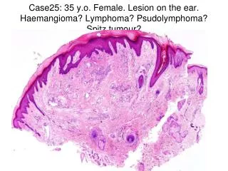
Case25: 35 y.o. Female. Lesion on the ear. Haemangioma? Lymphoma? Psudolymphoma? Spitz tumour?
CASE 25 - RC • Haemangioma • Angiolymphoid hyperplasia with eosinophilia • Kaposi’s sarcoma • Fibroma • Scar
CASE 25 - RC • Haemangioma • Angiolymphoid hyperplasia with eosinophilia • Kaposi’s sarcoma • Fibroma • Scar
Past Medical History Asthma Hayfever Allergy to cats and dogs Urticaria Drug History Loratidine Cetirizine Neoclarityn Levocetirizine Salbutamol inh Allergy to penicillin
FBC Eosinophils: 0.39 IgE 1667 (0-120) U&Es, LFTs, ESR normal MRI head No arteriovenous malformation Investigations
ALHE • Aetiology • Unknown • ?arteriovenous shunts • Gender • Females > males • Age • 30-50 y.o. • Site • Head and neck • Morphology • Dermal papules, nodules, plaques • Subcutaneous nodules Br J Derm 1969, 81:1-15, Br J Derm 1969, 81:804-816, J Am Acad Dermatol 1985, 12:781-796
Differential Diagnoses • Inflammatory angiomatous nodules • Pyogenic granuloma • Histiocytoid hemangioma • Kaposi’s sarcoma • Pseudolymphoma (lymphoid hyperplasia) • Cutaneous lymphoma • Kimura disease
Surgical excision Cryotherapy Curettage and Electrodessication Laser (argon, carbon dioxide, PDL, copper vapor) Radiotherapy Tacrolimus 0.1% ointment Intralesional corticosteroids Oral retinoids Topical imiquimod Photodynamic therapy Intralesional bleomycin Treatment of ALHE Dermatol Surg 2005, 31:713-16, J Dermatolog Treat 2004, 15(5):328-330, Dermatol Surg 2004, 30: 1169-1173, Dermatology 1998, 197: 189-191, J Am Acad Dermatol. Aug 2003;49 (2 Suppl Case Reports): S195-6, Clin Exp Dermatol 2009, June 17: Epub…..ahead of print
Management in our patient • ‘08: Nd:YAG laser • ‘08, ‘09: Tacrolimus 0.1% ointment • ‘09: electrodessication • ’09: Surgical excision • ’09: Topical Fluocinolone acetonide • ’09: Intralesional triamcinolone (40mg/1ml)