Download
1 / 106
1.11k likes | 1.27k Views
Understand the functions of the kidneys, anatomy of the urinary system, and kidney function overview. Learn about filtration, reabsorption, secretion, excretion, and micturition. Explore nephron types, tubular elements, and renal circulation.

E N D
RENAL PHYSIOLOGY dr. Adelia Handoko, M.Si PHYSIOLOGY DEPARTMENT UNIVERSITY OF JEMBER
Learning Objective • Functions of the Kidneys • Anatomy of the Urinary System • Overview of Kidney Function • Filtration • Reabsorption • Secretion • Excretion • Micturition
FUNCTIONS OF THE KIDNEYS • Regulation of extracellular fluid volume and blood pressure • Regulation of osmolarity. • Maintenance of ion balance • Homeostatic regulation of pH • Excretion of wastes • Production of hormones
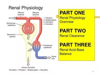
The Nephron Is theFunctional Unit of the Kidney • Vascular Elements of the Kidney • Blood flows from the afferent arteriole into a ball-like network of capillaries known as the glomerulus [glomus, a ball-shaped mass; plural glomeruli ] • Blood leaving the glomerulus flows into an efferent arteriole, then into a second set of capillaries, the peritubular capillaries [peri-, around] that surround the tubule • In juxtamedullary nephrons, the long peritubular capillaries that dip into the medulla are called the vasa recta
Nephron Type : • Cortical Nephrons • 85 percent of all nephrons • Located mostly within superficial cortex of kidney • Nephron loop (Loop of Henle) is relatively short • Efferent arteriole delivers blood to a network of peritubular capillaries • Juxtamedullary Nephrons • 15 percent of nephrons • Nephron loops extend deep into medulla • Peritubular capillaries connect to vasa recta
Tubular Elements of the Kidney • The combination of glomerulus and Bowman s capsule is called the renal corpuscle • Proximal tubule • Loop of henle • Descending limb • Ascending limb • Distal tubule • Collecting duct
Cortical radiate veins Cortical radiate arteries Interlobar arteries Cortex Segmental artery Adrenal artery Renal artery Renal vein Arcuate veins Interlobar veins Medulla Arcuate arteries A sectional view, showing major arteries and veins a
Glomerulus Cortical radiate vein Afferent arterioles Cortical radiate artery Arcuate artery Cortical nephron Arcuate vein Juxtamedullary nephron Renal pyramid Interlobar vein Interlobar artery Minor calyx Circulation in a single kidney lobe b
Renal vein Renal artery Segmental arteries Figure 26-5c The Blood Supply to the Kidneys. Interlobar arteries Interlobar veins Arcuate arteries Arcuate veins Cortical radiate veins Cortical radiate arteries Afferent arterioles Venules NEPHRONS Peritubular capillaries Glomerulus Efferent arteriole A flowchart of renal circulation c
Blood flow every 100 g tissue: ORGAN BLOOD FLOW ( ml / menit ) Skeletal muscle 3 Brain 54 Liver 58 Hearth muscle 84 Renal 420
Blood Supply to the Kidneys Renal Blood Flow (RBF) = 1200 ml/menit Cardiac Output (CO) = 5000 ml/menit RENAL FRACTION: = Part of total cardiac output that pass renal = Renal Blood Flow X 100% Cardiac Output • Kidneys receive 20–25 percent of total cardiac output
GLOMERULUS Filter Glomerulus: • Endotelium kapiler • Lamina basalis • Epitelium pars viseralis kapsula Bowman (Podocyt) Pori endotel = 100 nm Celah podocyt = 25 nm Lamina Basalis = 8 nm Luas area filtrasi = 0,8 nm² Ultrafiltrat = Plasma – Protein
NEPHRON Proximal convoluted tubule Distal convoluted tubule • Secretion of ions, acids, drugs, toxins • Variable reabsorption of water, sodium ions, and calcium ions (under hormonal control) • Reabsorption of water, ions, and all organic nutrients Figure 26-6 The Functional Anatomy of a Representative Nephron and the Collecting System (Part 1 of 2). Cuboidal cells with abundant microvilli Cuboidal cells with few microvilli Mitochondria Renal tubule Renal corpuscle • Production of filtrate Squamous cells Efferent arteriole Afferent arteriole Glomerulus Glomerular capsule Ascending limb of loop ends Descending limb of loop begins Capsular space Nephron loop Descending limb Further reabsorption of water Thick ascending limb Squamous cells Ascending limb Reabsorption of sodium and chloride ions Thin descending limb Low cuboidal cells KEY Solute reabsorption or secretion Filtrate Water reabsorption Variable solute reabsorption or secretion Variable water reabsorption
Table 26-1 The Organization of the Nephron and Collecting System (Part 1 of 2).
1. FILTRATION • The filtration of plasma into the kidney tubule is the first step in urine formation • The percentage of total plasma volume that filters into the tubule is called the filtration fraction
The Renal Corpuscle Contains Filtration Barriers • Substances leaving the plasma must pass through three filtration barriers before entering the tubule lumen: • glomerular capillary endothelium, • a basal lamina (basement membrane), • and the epithelium of Bowman’s capsule podosit
Capillary Pressure Causes Filtration • The three pressures: • capillary blood pressure • capillarycolloid osmotic pressure, and • capsule fluid pressure that influence glomerular filtration are summarize d
GFR is influenced by two factors: • Filtration pressure is determined primarily by renal blood flow and blood pressure. • The filtration coefficient has two components: • the surface area of the glomerular capillaries available for filtration and • the permeability of interface between the capillary and Bowman’s capsule. • In this respect, glomerular filtration is similar to gas exchange at the alveoli, where the rate of gas exchange depends on partial pressure differences, the surface area of the alveoli, and the permeability of the alveolar membrane.
GFR Is Relatively Constant • The volume of fluid that filters into Bowman s capsule perunit time is the glomerular filtration rate (GFR). • Average GFR is 125 mL/min, or 180 L/day • Blood pressure provides the hydrostatic pressure that drives glomerular filtration. Therefore, it might seem reasonable to assume that if blood pressure increased, GFR would increase, and if blood pressure fell, GFR would decrease. • That is not usually the case, however. Instead, GFR is remarkably constant over a wide range of blood pressures. • As long as mean arterial blood pressure remains between 80 mm Hg and 180 mm Hg, GFR averages 180 L/day • GFR is controlled primarily by regulation of blood flow through the renal arterioles
GFR Is Subject to Autoregulation • Autoregulation of glomerular filtration rate is a local control process in which the kidney maintains a relatively constant GFR in the face of normal fluctuations in blood pressure. • One important function of GFR autoregulation is to protect the filtration barriers from high blood pressures that might damage them.
We do not completely understand the autoregulation process, but several mechanisms are at work : • The myogenic response is the intrinsic ability of vascular smooth muscle to respond to pressure changes. • Tubuloglomerular feedback is a paracrine signaling mechanism through which changes in fluid flow through the loop of Henle influence GFR
Autoregulation Immediate local response in the kidney HOMEOSTASIS RESTORED Increased glomerular blood pressure if sufficient Normal GFR Dilation of afferent arterioles HOMEOSTASIS DISTURBED HOMEOSTASIS Normal glomerular filtration rate Decreased GFR resulting in decreased filtrate and urine production Contraction of mesangial cells Start Constriction of efferent arterioles
Hormones and Autonomic Neurons Also Influence GFR • Neural control of GFR is mediated by sympathetic neurons that innervate both the afferent and efferent arterioles. Sympathetic innervation of α-receptors on vascular smooth muscle causes vasoconstriction • A variety of hormones also influence arteriolar resistance. Among the most important are angiotensin II, a potent vasoconstrictor, and prostaglandins, which act as vasodilators.
Renin–Angiotensin-Aldosterone System Integrated endocrine and neural mechanisms activated Renin in the bloodstream triggers formation of angiotensin I, which is then activated to angiotensin II by angiotensin converting enzyme (ACE) in the capillaries of the lungs Endocrine response Figure 26-11 The Response to a Reduction in the GFR (Part 1 of 2). Juxtaglomerular complex increases production of renin Angiotensin II triggers increased aldosterone secretion by the adrenal glands Angiotensin II triggers neural responses Angiotensin II constricts peripheral arterioles and further constricts the efferent arterioles Aldosterone increases Na+ retention HOMEOSTASIS RESTORED Increased stimulation of thirst centers Increased fluid consumption Increased systemic blood pressure Increased blood volume Increased glomerular pressure Increased ADH production Increased fluid retention Constriction of venous reservoirs Increased sympathetic motor tone Increased cardiac output Together, angiotensin II and sympathetic activation stimulate peripheral vasoconstriction HOMEOSTASIS Normal glomerular filtration rate
2. Reabsorption • Each day, 180 liters of filtered fluid pass from the glomerular capillaries into the tubules, yet only about 1.5 liters are excreted in the urine. • Thus more than 99% of the fluid entering the tubulesmust be reabsorbed into the blood as filtrate moves through the nephrons. • Most of this reabsorption takes place in the proximaltubule, with a smaller amount of reabsorption in the distal segments of the nephrons.
Reabsorption May Be Active or Passive • Reabsorption involves both : • epithelial transport (also called transcellular transport), in which substances cross both the apical and basolateral membranes of the tubule epithelial cell • paracellular pathway, in which sub stances pass through the junction between two adjacent cells
Active Transport of Sodium • The active transport of Na+ is the primary driving force for most renal reabsorption