Download
1 / 88
1.05k likes | 2.1k Views
Lymphatic tissue. Components of lymphoid system. Lymph and lymph nodes Lymph – fluid scavenged from the intersitium Collected in thin walled lymphatic vessels Returned to general circulation Contains leukocytes Filtered through small specialized organs (lymph nodes). Lymph.

E N D
Components of lymphoid system • Lymph and lymph nodes • Lymph – fluid scavenged from the intersitium • Collected in thin walled lymphatic vessels • Returned to general circulation • Contains leukocytes • Filtered through small specialized organs (lymph nodes)
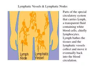
Lymph • Capillaries lose 2-4 L of fluid and plasma protein • Lymphatic system absorbs excess fluid and returns it to the bloodstream • Makes sure circulatory system has enough blood to work properly • Decreases edema
Lymph– • Clear colorless fluid—like plasma • Less proteins • Has bacteria, virus, cell debris • Lipid content high • Lymphocytes content high • Many in capillary bed • Lymphatic vessels • Take fluid from tissue back to the bloodstream—veins • Usually along side of vein • Similar to vein (low pressure, use SK muscle and respiratory pump to help flow, valves also)
Lymphoid Cells • The two main varieties are T cells and B cells • Both types originate from stem cells in bone marrow • T cells – cell mediated immunity • B cells – humoral/ antibody mediated immunity • Small and large lymphocytes in peripheral blood • Large are the activated B lymphocytes • Antigen – anything the body perceives as foreign • Bacteria and their toxins; viruses • Mismatched RBCs or cancer cells
Lymphocytes • T cells • Manage the immune response • Attack and destroy foreign cells • B cells • Produce plasma cells, which secrete antibodies • Plasma cells are derived from activated B lymphocytes that have left the blood stream and taken up residence in connective tissue. SPOKE wheel appearance • Antibodies immobilize antigens
Other Lymphoid Cells • Macrophages – phagocytize foreign substances and help activate T cells • Dendritic cells – spiny-looking cells with functions similar to macrophages • Reticular cells – fibroblast like cells that produce a stroma, or network, that supports other cell types in lymphoid organs
Lymphoid Tissue • Lymphatic follicles (nodules) • Solid, spherical bodies consisting of tightly packed reticular elements and cells • Have a germinal center composed of dendritic and B cells • Found in isolation and as part of larger lymphoid organs
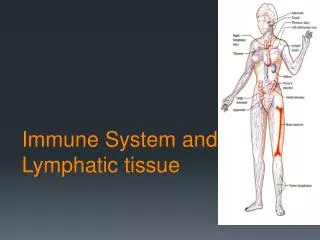
Introduction • Lymphoid system consists of cells, tissues & organs that contain aggregates of lymphocytes. The lymphoid organs are: • Primary lymphoid organs – bone marrow & thymus • Secondary lymphoid organs – lymph nodes, spleen, tonsils, Peyer’s patches, MALT (mucosa associated lymphoid tissue)
Lymphoid Organs Central Origin and main differentiation Thymus Bone marrow Spleen Lymphoid nodes Solitary nodules Tonsils Appendix Peyer’s patches Peripheral Proliferation and additional differentiation Immune response
Lymphoid Organs • Encapsulated • Thymus • Lymph nodes • Spleen • Unencapsulated • Diffuse • Tonsils
Thymus • A bilobed organ that secrets hormones (thymosin and thymopoietin) that cause T lymphocytes to become immunocompetent • The size of the thymus varies with age • In infants, it is found in the inferior neck and extends into the mediastinum where it partially overlies the heart • It increases in size and is most active during childhood • It stops growing during adolescence and then gradually atrophies
Internal Anatomy of the Thymus • Thymic lobes contain an outer cortex and inner medulla • The cortex contains densely packed lymphocytes and scattered macrophages • The medulla contains fewer lymphocytes and thymic (Hassall’s) corpuscles
Thymus • The thymus differs from other lymphoid organs in important ways • It functions strictly in T lymphocyte maturation • It does not directly fight antigens • Function: • production of immunocompetent T lymphocytes • production of mature but naïve T cells for peripheral tissues and circulation • immunological self-tolerance • regulation of T cell maturation, proliferation and function via secretion of hormones
Thymic Cortex • Consists of proliferating lymphocytes dispersed among macrophages & epithelial reticular cells. • These epithelial cells/ reticular cells form the support structure for the developing T cells but also play an important role in isolating the T cells from foreign anitgens during their development • They are of endodermal origin& do not secrete reticular fibers.
Epithelial Reticular Cells • Endodermally derived. • Stellateeosinophilic cells with an oval nucleus. • Joined to adjacent reticular cells by desmosomes. • Enclose the developing lymphocytes within them. • Help in the formation of a blood-thymus barrier - prevents antigenic stimulation of the lymphocytes & allows for proper differentiation. • Secrete various hormones (thymulin, thymopoietin, thymosin & thymichumoral factor) that regulate T cell differentiation.
Blood Thymus barrier • Prevents premature exposure of lymphocytes to antigen (to inhibit tolerance). • The integrity of the space within the epithelial cell framework is extremely important because it prevents the premature stimulation of T cells by antigens • Non-fenestrated endothelium with occluding junctions (major component of the barrier) • Endothelium • Thick basal lamina • Pericytes • Macrophages • Type I epithelioreticular cells
Thymic Cortex • Contains maturing lymphocytes, macrophages & reticular cells. • Many lymphocytes die by apoptosis & are removed by macrophages. • Outer cortex is populated by immature lymphocytes & as they mature they migrate to the deeper part of the cortex.
MHC I Immature T cell Immature T cell Immature T cell CD4 CD4 CD4 CD8 CD8 CD8 Epithelio reticular cell (RE) MHC II MHC I T cells that recognize MHC I are destroyed or they lose CD4 from their surface CD4 +ve CD8 +ve T cell T cell MHC II Maturation of T cell in thymus CORTEX • First T cell has to acquire markers CD4 and CD8 • Cell without marker is destroyed RE cell teaches the immature T cell to recognize the MHC markers Some identify MHC I and some MHC II T cell who don’t learn to identify either are destroyed MEDULLA
Thymic Medulla • Hassall’s corpuscles concentrically arranged, flattened epithelial reticular cells that become filled with keratin filaments, degenerate & may become calcified. • They appear in the fetus & their number gradually increases after birth.
QUESTION The production of new T lymphocytes in the thymus occurs in which of the following regions? • Superficial cortex • Corticomedullary junction • Thymic nodules • Deep medulla • Thymic corpuscles
Lymph Node • A filter: • purifies lymph before return to venous circulation • Removes: • debris • pathogens • 99% of antigens • Initiation of immune response • Activation & proliferation of lymphocytes
Function of lymph nodes • filter debris and microorganisms via phagocytosis by fixed macrophages • facilitate the interaction between antigen presenting cells and circulating lymphocytes to initiate an immune response • B lymphocytes: activation and proliferation; plasma cell formation and antibody production in response to antigens • T lymphocytes: activation to become T helper and T cytotoxic cells
Lymphnode structure • Afferent vessels – convey lymph towards the node • Efferent vessels – drain lymph away from the node at the hilum • Capsule • Trabeculae • Reticular tissue
Lymph node Capsule Subcapsular sinus
Structure of a Lymph Node (Cortex) • Superficial cortex: The cortex contains follicles with germinal centers, heavy with dividing B cells • Dendritic cells nearly encapsulate the follicles • Deep cortex houses T cells in transit • T cells circulate continuously among the blood, lymph nodes, and lymphatic stream
Lymphoid follicle • Germinal Center (GC) - contains pale-staining cells. The open, pale-staining nature of the nuclei of these cells indicate that they are B lymphocytes undergoing active proliferation. • Other cells include: • Follicular dendritic cells that present antigen to the B cells • Macrophages that engulfed dead B cells that have died by apotosis
Lymphoid follicle cortex • Germinal center - Functional aspects: • resting B cells enter the lymph node parenchyma though the high endothelial venules and if they encounter an antigen with which they can react, they then enter the cycle of blast transformation to produce clones of plasma cells and B memory cells. • This production of clones occurs in the germinal centers of lymphoid follicles. • Mantle zone (corona) – • The germinal center is surrounded by a ring of darker-staining cells. The condensed nature of their nuclei indicates that these are resting B cells. Also present in the mantle zone are T helper cells, macrophages and dendritic cells.
Paracortical zone / deep cortex • Paracortical zone - deeper regions of the cortex contain primarily T lymphocytes that do not form into follicles. • T lymphocytes (helper and cytotoxic/suppresor) arrive through the circulation, enter the lymph node parenchyma through the high endothelial venulesand take up residence in the paracortical zone. • If activated, the T lymphocytes undergo active proliferation to produce expanded clones of activated T lymphocytes. • Functional aspects: • T lymphocytes that arrive at the lymph node via the arterial blood stream gain access to the parenchyma of the lymph node through the wall of the high endothelial venules located in the paracortical zone. • These blood vessels contain endothelial cells that are expressing specific lymphocyte binding molecules called addressins. • These surface molecules are available to bind to lymphocytes that recognize them, the lymphoctyes bind to the surface of the endothelium, then cross the vessel wall and enter the lymph node parenchyma.
Homing of lymphocytes • A process by which lymphocytes return to lymph node by crossing walls of blood vessels, High endothelial venules- present in lymph nodes.
Lymph node architecture • HEV – specializedPost-capillary venules. • Cuboidal endothelium • Concentrates lymph • Receptors for antigenprimed lymphocytes *HEV- High endothelial venules
Structure of a Lymph Node (Medulla) • Medullary cords extend from the cortex and contain B cells, T cells, and plasma cells • The medullary cords are composed of plasma cells producing antibodies, their precursors, macrophages and T helper cells. • The most prominent cell in the cord is the precursor to plasma cells or immunoblasts that came from the germinal centers of the lymphoid follicles in the cortex of the node. • In the medullary cords, the plasma cells undergo final maturation and secrete antibodies into the lymph that is collected by efferent lymphatic vessels in the node and eventually carried to the general circulation. Plasma cells may also get into the general circulation in this manner
Structure of a Lymph Node (Medulla) • The medullary sinuses are composed primarily of • reticular fibers (RF) providing the support framework, • reticular cells (fibroblast-like cells that secrete the reticulin) (RC) and • macrophages.
Lymph node • Cell types • Lymphocytes and plasma cells • Accessory cells • Macrophages • Follicular dendritic cells (FDC) • Interdigitating dendritic cells (DC) • Stromal cells
Lymphatic Disorders: • Elephantiasis: parasite, roundworm • Lives in lymph nodes • Prevents fluid flow back to circulatory system • Chronic edema (fluid swelling) • Leads to thickening of skin • Autoimmune diseases • Failure of self tolerance • Immune system fails to recognize self antigens from foreign ones and attacks body’s own tissues e.gLupus • Young women more impacted usually • Autoimmune disease against DNA