Download
1 / 1
40 likes | 422 Views
Though partial overlaps between mandibular third molars and mandibular canals are detected in panoramic radiograph, there may be many cases of encroachment of the canal. Therefore, to define a more accurate relationship between the two, we require CBCT or CT images.

E N D
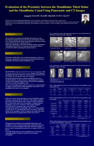
Though partial overlaps between mandibular third molars and mandibular canals are detected in panoramic radiograph, there may be many cases of encroachment of the canal. Therefore, to define a more accurate relationship between the two, we require CBCT or CT images. The results of this study may lead to guidelines for oral surgeons evaluating whether to obtain an axial CT scan for further investigation. 1) point contact with canal 2) superimposition 3) partially overlapped Evaluation of the Proximity between the Mandibular Third Molar and the Mandibular Canal Using Panoramic and CT Images Joung JI1, Park SH1, Choi BR1, Huh KH2, Yi WJ3, Choi SC2 1Department of Oral and Maxillofacial Radiology, School of Dentistry, Seoul National University, Seoul, South Korea , 2Department of Oral and Maxillofacial Radiology, Dental Research Institute, School of Dentistry, Seoul National University, Seoul, South Korea, 3Department of Oral and Maxillofacial Radiology, Dental Research Institute and BK21, School of Dentistry, Seoul National University, Seoul, South Korea. Fig 1. Classification of the relationship between the root of lower third molar and mandibular canal in panoramic radiograph. Introduction The extraction of an impacted mandibular third molar can cause neurological complications due to the damage in the inferior alveolar nerve. It is known that the risk increases markedly when there is direct contact between the nerve and the tooth root. Panoramic radiographs are the most commonly used for assessing the relationship between these two structures preoperatively. But it is restrictively useful for predicting the physical contact. Objective The purpose of this study was to evaluate the diagnostic accuracy of panoramic radiograph in assessing the proximity between the root of mandibular third molar and the mandibular canal. 4) interruption of the border 5) diversion of mandibular canal 6) narrowing of mandibular canal Fig 2. Classification of the relationship between the root of lower third molar and mandibular canal in CT image Materials &Method The panoramic [Orthopantomograph op100 (Instrumentarium Corp., Tuusula, Finland)] and CT [Somatom Sensation 10 (Siemens AG, Forchheim, Germany) ] images of 385 impacted mandibular third molar which were obtained from 235 patients (male: 113, female: 125, average age: 28.6) were evaluated. The proximity between the root and the mandibular canal were classified as follows; 1) point contact, 2) superimposition, 3) partial overlap, 4) interruption of upper border, 5) diversion of mandibular canal, 6) narrowing of mandibular canal. The actual relationship between the two structures was assessed using CT images. Also the location of the mandibular canal relative to the mandibular third molar was investigated. 1) no contact 2) between inner wall and outer wall of mandibular canal 3) blurred inner wall 4) narrowing of the mandibular canal 5) encroaching the mandibular canal Table 1. Relationship between the root of lower third molar and mandibular canal in panoramic view Results Table 2. Relationship between the root of lower third molar and mandibular canal in CT images In the case of roots' being superimposed on the mandibular canal in panoramic radiograph, 49.6% and 13.9% of the samples exhibited narrowing and encroachment of the canal in CT image respectively. On the other hand, when roots were partially overlapped with the mandibular canal, 23.6% and 22.4% of the samples exhibited narrowing and encroachment of the canal respectively. Considering the position of mandibular canal, 35.7% of the samples were buccal, 25.7% were lingual and 33.6% were inferior. When the position of the mandibular canal was lingual, narrowing of the canal appeared much more frequently than in other cases (67.0%). * MC: mandibular canal, ( ): % Conclusion & discussion Table 3. Relationship between the root of lower third molar and mandibular canal according to the mandibular canal position * MC: mandibular canal, ( ): %