Download
1 / 79
800 likes | 1.06k Views
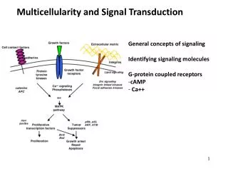
Multicellularity and Signal Transduction. General concepts of signaling Identifying signaling molecules G-protein coupled receptors cAMP Ca++. 1. Multicellularity. Cellular Specialization Separation of functions Different lineages of cells Established in development

E N D
Multicellularity and Signal Transduction • General concepts of signaling • Identifying signaling molecules • G-protein coupled receptors • cAMP • Ca++ 1
Multicellularity Cellular Specialization Separation of functions Different lineages of cells Established in development Ectoderm- ex. neurons, epithelium Mesoderm- ex. muscle, blood Endoderm- ex. gut, liver Maintenance in adulthood Tissue-specific stem cells proliferation and differentiation 2
epinephrine Adrenaline/epinephrine in blood stream alters physiology Cellular components translate signal into action Sensing Environmental Signals Ex. “Fight or Flight” Response 3
Cellular components translate signal into action Cell Communication • Cells have the ability to communicate with each other • Signaling molecules function within an organism to control: • Allows cells to respond to many environmental signals • Signaling molecules are synthesized and released from certain cells
Molecular Components that Sense and Respond to External Stimuli External signal- ligand Signal receiver- receptor in plasma membrane Messengers- intracellular components that communicate signal Effectors- intracellular players that mediate response to signal 5
Signal Transduction • General Steps: • Synthesis of signaling molecules by signaling cell • Release of signaling molecule • Transport of signal molecule to target cell • Binding of signal molecule to receptor on target cell • Initiation of intracellular signal transduction • Specific changes to cellular functions • Feedback regulation 6
Fast vs. Slow Responses Signaling Cell Signal sent from cell or environment Target cell -expresses receptor that binds to signaling molecule -contain intracellular proteins that transmit signal -changes occur in target cell fast response slow response Receiving Cell 7
Cellular Response to Signaling • (1) changes in protein activity or function • (2) changes in the amounts of specific proteins • The first type of response occurs more rapidly than the second • Changes in the activity of pre-existing proteins
Cellular Response to Signaling • Several intracellular proteins or small molecules contribute to signaling: • Cytosolic enzymes • Ligand binding to a receptor • G proteins • Small molecules
First and Second Messengers • Binding of ligands (the first messengers) to cell-surface receptors leads: • Binding of ligands can also cause a decrease in concentration of the second messenger
Receptors • Most signaling molecules are too large and/or hydrophobic to get through the cell membrane • Need protein receptors
Signaling • Rapid responses to the environment are primarily mediated by the nervous system and by hormones • Cells that generate these signals are found in the pancreas, pituitary glands, neurons, hypothalamus
Signaling • Exocytosis of the stored molecules • Released molecules only last for seconds or a few minutes • Short term responses • Released signaling molecules travel to target cells
Signaling • Synthesis and • Release of the signaling molecule • Transport of the signal to the target cell • Binding of the signal by a specific receptor • Initiation intracellular signal-transduction pathways • Specific changes in cellular function, metabolism, or development • Deactivation of the receptor • Removal of the signaling molecule to terminate response
Signaling • Endocrine: signaling molecules are synthesized and secreted by signaling cells (endocrine cells) • Autocrine: cells respond to substances that they release themselves
Signaling • Paracrine: signaling molecule released by a cell affect only nearby cells
Synthesis, Release and Transport of Signal: Endocrine Secreted signals Endocrine: Long distance signal Hormone secretion into blood stream 17
Synthesis, Release and Transport of Signal: Paracrine and Autocrine Secreted signals Paracrine: Short distance signal Signaling molecule secreting from cell adjacent to target cell Autocrine: Self signal Target cell secretes its own signal 21
Signaling • Membrane bound signals on one cell bind to receptors on adjacent cells to trigger differentiation • Some can act in short and long ranges
Signal between adjacent, attached cells Cell-cell contact Signaling cell expresses membrane-bound ligand and target cell expresses membrane-bound receptor 23
Strategies to Identify Signaling Components • Biochemical • Molecular • Genetic 24
Identification and purification of a receptor Affinity Chromatography Couple ligand to bead and load Column Run receptor-containing extract over Column Receptor binds to ligand on beads Wash and discard flow through Add free ligand in excess Receptor binds to free ligand Collect ligand-receptor complexes Sequence peptide Isolate gene Express putative receptor gene and test binding of ligand Biochemically 25
Identification and purification of a receptor Molecular cloning Identify cells lacking receptor; do not bind ligand and do not respond Introduce collection of cDNA isolated from cell that do express receptor and respond to signal Add ligand X and look for clones of cells that do respond to ligand Isolate cDNA that confers response Sequence Confirm by reintroducing putative receptor cDNA into cells and retesting 26
X Genetic Isolation of signaling molecules B cone B cone B R7 B R7 A A A Define a cellular output for activation of receptor B cell becomes R7 neuron when signal detected Signal Target Selective killing of A cell results in B cell becoming a cone cell instead of R7 neuron A cell sends signal to B cell to become R7 neuron Perform genetic screen for loss of cellular output that resembles loss of signaling cell B cell becomes cone cell instead of R7 neuron Loss of signal can be from mutation in ligand, receptor, or intracellular effectors 27
GPCRs • Most common class of receptors is called G Protein-Coupled Receptors (GPCRs) • About 900 • Activation of these receptors alters gene expression • Many different receptors, one ligand
GPCR signaling in different tissues can elicit different responses epinephrine Increase in cAMP levels Glycogen breakdown Glucose release Increase in intracellular Ca++ Muscle Contraction 30
GPCRs • G protein coupled receptor ligand binding triggers the G protein to exchange GDP for GTP 29
Cellular Response to Signaling • Two large classes of GTPase switch proteins are used in signaling. • Trimeric • Monomeric
G-protein coupled receptors 7-pass transmembrane receptors N-term extracellular C-term cytoplasmic Associate with trimeric G-proteins Act as GEF for G-protein Lead to increases in second messengers cAMP Ca++ Effector molecules found in membrane 32
GPCRs • All GPCRs have the same orientation in the membrane 33
GPCRs • Exterior surface consists mainly of hydrophobic amino acids
GPCRs • TrimericG proteins have three subunits, α, β and γ • During signaling, the β and γ subunits remain together • Without ligand, the α subunit is bound to GDP
Trimeric G-protein Composed of 3 subunits: α, β, and γ α contains GTPase activity α and γ tethered to plasma membrane via lipid tails When α bound to GDP, also interacts with β and γ−−> This is the off position When GPCR activated, binds to G-protein and acts as a GEF Stimulates exchange of GDP for GTP in the α subunit 36
GPCR signaling • Ligand binding causes a conformational change, • When GTP binds, another conformation change • Usually, Gα remains anchored to the membrane • Gα-GTP can inhibit the effector • Gβγ is sometimes freed from α to signal through an effector protein • GTP is hydrolyzed by α, which has GTPase activity • This switches α back to the GDP state and prevents any further activity • Binding of α to the effector protein can also cause hydrolysis • It then quickly reassociates with βγ, ready to repeat the process
GPCRs that regulate ion channels • Acetylcholine receptors in the heart muscle activate a G protein that opens K+ channels • Ligand binding leads to the opening of the associated K+ channels
GPCRs that regulate Adenylate Cyclase • Adenylatecyclase is a very common effector protein of GPCRs • When blood sugar levels are low, cells have a demand for glucose • In the liver, these two bind different GPCRs, but the response is the same • Activation and inhibition of cAMP occurs though many different ways and in different cell types
GPCRs that regulate Adenylate Cyclase • Binding of prostaglandin PGE1 or adenosine to their GPCRs inhibits adenylatecyclase
Adenylyl (Adenylate) Cyclase Enzyme that converts ATP to cAMP Activated by Gαs Intergral membrane protein 45
GPCRs that regulate Adenylate Cyclase • cAMP activates Protein Kinases A by releasing catalytic subunits • Inactive PKA is a tetramer that has two regulatory (R) subunits and two catalytic (C) subunits • Inactive PKA is turned on by cAMP, as each R subunit has a cAMP binding site (CNB-A and CNB-B)
Epinephrine • There are different receptors for epinephrine in many different types of cells • Mediates the body’s response to stress